Quantcast
Channel: Guérir du Cancer

Entropy as the Driving Force of Pathogenesis: an Attempt of Classification of the Diseases Based on the Laws of Physics

$
0
0

Chers tous,
Si j’ai choisi le silence médiatique dans la période difficile que nous avons tous traversée, c’est pour avancer sur le plan théorique et pratique pour un traitement efficace du cancer. Ce papier technique est le premier d’une série qui tend à démontrer que le cancer n’est pas une maladie particulière. Le cancer est causé par l’effet Warburg, lui même une conséquence du deuxième principe de la thermodynamique. Comprendre que le cancer s’inscrit de façon, somme toute banale, dans les lois de la physique nous permettra, je l’espère d’améliorer le traitement métabolique.

À tous
Laurent


Lire le document (en anglais)

L’article Entropy as the Driving Force of Pathogenesis: an Attempt of Classification of the Diseases Based on the Laws of Physics est apparu en premier sur Guérir du Cancer.


Targeting Mitochondrial Singlet Oxygen Dynamics Offers New Perspectives for Effective Metabolic Therapies of Cancer

$
0
0


Le cancer croit et se développe car la tumeur fermente. C’est l’effet Warburg. Nous le ciblons avec des molécules qui modifient le métabolisme du sucre. Mais pour que le sucre brûle, il faut que l’oxygène soit actif. Hors l’oxygène existe sous deux formes quantiques. S’il est sous forme triplet comme l’oxygène que vous respirez il est inactif. Il est activé par la mitochondrie en oxygène singulet qui lui peut bruler les dérivés du sucre. Si cette activation d’une forme inerte en une forme active ne se fait pas, il y aura l’effet Warburg. Il est probable que l’effet Warburg et donc le cancer soit la conséquence de cette inactivation de l’oxygène. La porte est ouverte vers de nouveaux traitements;
Laurent


 Jorgelindo da Veiga Moreira1*,  Laurent Schwartz2 and  Mario Jolicoeur1
  • 1Research Laboratory in Applied Metabolic Engineering, Department of Chemical Engineering, Polytechnique Montréal, Montréal, QC, Canada
  • 2Assistance Publique des Hôpitaux de Paris, Paris, France
The occurrence of mitochondrial respiration has allowed evolution toward more complex and advanced life forms. However, its dysfunction is now also seen as the most probable cause of one of the biggest scourges in human health, cancer. Conventional cancer treatments such as chemotherapy, which mainly focus on disrupting the cell division process, have shown being effective in the attenuation of various cancers but also showing significant limits as well as serious sides effects. Indeed, the idea that cancer is a metabolic disease with mitochondria as the central site of the pathology is now emerging, and we provide here a review supporting this “novel” hypothesis re-actualizing past century Otto Warburg’s thoughts. Our conclusion, while integrating literature, is that mitochondrial activity and, in particular, the activity of cytochrome c oxidase, complex IV of the ETC, plays a fundamental role in the effectiveness or non-effectiveness of chemotherapy, immunotherapy and probably radiotherapy treatments. We therefore propose that cancer cells mitochondrial singlet oxygen (1O2) dynamics may be an efficient target for metabolic therapy development.
 

Introduction

Oxygen is “the molecule that made the world” stated Lane in his seminal book (1). Nowadays, the atmosphere of Earth is composed of 21% oxygen and used by most organisms for respiration (animals, plants, and many prokaryotes). These living organisms use oxygen as oxidizing agents to retrieve energy from reduced compounds (2).

Human body is made up of 10,000–100,000 billion cells. Every day tens thousands of cancer cells are formed but are also soon eliminated by the immune system (3). The other common point between cancer cells and pathogenic organisms is their dependence on metabolism of healthy cells and the irrigation of nutrients by the blood system to ensure their replication (4). In this way, competition arises between cancer cells and somatic cells for access to nutrients, specially to glucose, and oxygen. This competition is similar to game theory as introduced by John von Neumann (5) then applied to biology by John Maynard Smith (6). Transposed to game theory, the metabolism of healthy cells and cancer cells are distinguished by the way they produce energy in the form of ATP. In presence of oxygen, healthy cells use glucose to produce about 30 molecules of mitochondrial oxidative phosphorylation ATP. Under hypoxic conditions, they produce only 2 molecules of ATP through glycolysis and release 2 molecules of lactic acid. As far as cancer cells are concerned, they favor the glycolysis pathway for energy production, even in oxygenated environment, referred as aerobic glycolysis or “Warburg effect” named after the German physician and biochemist Otto Heinrich Warburg who first reported this observation (79). Warburg showed that cancer cells in culture have higher rates of glucose consumption and lactate secretion compared to normal cells and hypothesized mitochondrial dysfunction to explain this glycolytic phenotype. However, many more recent studies have demonstrated the integrity of mitochondria in cancer cells (10, 11).

Here, we bring an overview of the common therapeutic approaches such as chemotherapy to circumvent tumor progression and limits thereof. We specially focused on the involvement of mitochondria regarding the metabolic adaptation of cancer cells to escape from apoptosis and promote, in some way, cancer recurrence (12). In perspective of this study, we propose that the cancer metabolic phenotype or Warburg effect could be relieved by the controlled generation of mitochondrial singlet oxygen (1O2). Singlet oxygen is the first excited state of the dioxygen molecule and is part of the ROS species generated during the OXPHOS process. We support the idea that the generation of 1O2 in the respiratory chain is a necessary step for OXPHOS and ATP production. Our hypothesis is that the mitochondrial accumulation of ROS observed during phases of high energy demand or in periods of substrates deficiency would be due to a limitation of the respiratory capacity of cells in general, and in particular in tumors where physical and metabolic variabilities have been reported (13). This, is a fundamental review deciphering on how cancer cells’ ROS production is an adaptation to escape apoptosis and how strategies such as singlet oxygen-oriented therapy could potentially counteract cell proliferation and metastasis.


Mitochondrial Respiration is the Key to Understand Cancer Metabolism

The cell cycle is also a metabolic cycle regulated by redox transitions promoting fermentation or cellular respiration (14). Cancerous masses deploy a panoply of adaptative actions in the face of external stresses such as chemotherapy. This is partly made possible by the “hijacking” of the genetic program for regulating the cell cycle. In the following, we will thus first describe the mitochondrial metabolism and the electron transfer chain (ETC) functioning. Then we will decipher how cancer cells manage to “hijack” mitochondrial activity in its favor and to escape from apoptosis even under ROS-induced chemotherapy treatments.

Energetic Metabolism and the Respiratory Chain

Mitochondria are involved in various cellular processes such as differentiation and cell death (apoptosis) as well as supporting the immune response (15, 16). The mitochondria are the energy factories in eukaryotes. They mobilize the enzymes necessary for the proper functioning of the citric acid cycle (TCA, or Krebs cycle) and are involved in the management of the redox balance. Mitochondria use oxygen to extract energy from carbon-based nutrients found in the cellular environment or stored as intracellular macromolecules like glycogen and results in the synthesis/recycling of adenosine triphosphate (ATP) by phosphorylation of adenosine diphosphate (ADP). This process called oxidative phosphorylation (OxPhos) takes place at the level of the mitochondrial ETC chain.

The “Classical” Respiratory Chain of Eukaryotic Cells

Peter D. Mitchell proposed in 1961 the chemiosmotic theory to explain the synthesis of ATP by OxPhos mechanism. This theory suggests that the production of ATP is made possible by a proton gradient (ΔpH) formed on either sides of the inner membrane of the mitochondria thanks to an ATPase catalyzing the reaction (17). The proteins animating this respiratory chain promote the creation of this ΔpH thanks to the energy of the redox couples (NAD+/NADH and FAD/FADH2) present in the mitochondrial matrix and brought about by the catabolism of carbon-based resources. ATPase takes advantage of this electrochemical gradient also called protonmotive force (Δp) for the phosphorylation of ADP into ATP. The ETC of eukaryotic cells is essentially composed of enzymes and co-enzymes involved in the transfer of electrons and the synthesis of ATP. Four protein complexes (CI-IV) are involved in electron transport and oxygen reduction. ATP synthase, also known as complex V (CV), catalyzes ATP synthesis (Figure 1) (1801). A special focus is placed on complex IV (IV) or cytochrome c oxidase, since this enzyme has a pivotal role in mitochondrial respiration. It catalyzes the transfer of electrons from the reduced form of cytochrome c to dioxygen, which becomes the final acceptor of electrons in the respiratory chain (19, 20). Two molecules of water are formed through this reaction and four protons are pumped to the intermembrane space. This enzyme has been well-studied, in particular for understanding the mechanisms allowing both the reduction of dioxygen into water molecules and the flow of protons through the protein structure (21). The pivotal role of prosthetic (22) groups has been demonstrated. These metallic prosthetic groups form two copper redox centers (CuA and CuB) and two heme centers (heme a and heme a3). CuB and heme a3 are physically close and form a bi-nuclear center where the oxidation of oxygen takes place. CuA is the first electron acceptor of cytochrome c while heme a serves as an intermediary for the transfer of electrons between CuA and the binuclear center for the reduction of O2 (18). A detailed model of the O2 activation and its reduction cycle has been proposed (23, 24).

 
FIGURE 1
www.frontiersin.org
Figure 1. The electron transfer chain (ETC). It is composed of four complexes involved in electron transfer and proton translocation. Complex V or ATP synthase catalyzes ATP synthesis. Uncoupling proteins and molecules are also reported to trigger proton leaks to mitochondrial matrix.

 
Regulated Steps of Mitochondrial Respiration

The desire to identify a major OXPHOS “controller” began in the 70–80s (25), notably thanks to contributions of the Metabolic Control Theory (MCT) (2628). CIV, CV, and the mitochondrial ATP/ADP translocase were the main candidates (29, 30). Subsequently, work on mitochondria isolated from liver cells has highlighted the role of these proteins, depending on the energy state of the cell (29). In short, during highly active OxPhos phase, the supply of electrons from substrates and the electron flux through the ETC were estimated as the limiting steps. During the stationary phase of respiration, proton translocation through mitochondrial membrane was the limiting step. Finally, when the cell’s energy demand is reduced, the activity of ATP translocase became the limiting factor for OxPhos.

The Potential Role of Apoproteins in Mitochondrial Mode Switch

ETC complexes behave like real biological capacitors, by analogy with electrical circuits. They have the capacity to store the electrons recovered from NADH or succinate. In 1964, John Rieske isolated a subunit of Complex III. He highlighted the presence of an iron-sulfur atomic aggregate on this apoprotein (31). This component of complex III has since become the Rieske protein while other proteins have been identified and assimilated to the Rieske protein due to the presence of the iron-sulfur cluster [Fe-S]. This is the case for complexes I and II of the respiratory chain (32, 33) but also for aconitase (34) which also consist of one or more several centers [Fe-S]. One may wonder if inhibition of these clusters could impair cellular respiration. Few studies have been devoted to this problem. Graham and Trumpowe (35) carried out mutations in the conserved domains of the Rieske protein in the yeast Saccharomyces cerevisiae. They observed that directed-mutations on codons of amino acids known to interact specifically with the [2Fe-2S] cluster and present exclusively in aerobic organisms lead to an inability of these mutants to use non-fermentable carbon sources for their growth (35). As described above, this prosthetic group, which is also present in cytochrome bc1 (complex III) is the first electron acceptor emitted by ubiquinol (Q) and participates in the creation of the protonmotive force. Other studies emphasize the importance of the [2Fe-2S] cluster in maintaining OxPhos. Diaz et al. (36) have notably demonstrated that mutations on the gene coding for Rieske protein in fibroblasts causes a decrease in the synthesis of complexes I and IV and OxPhos deficiency (36). These [Fe-S] clusters are not the only prosthetic groups to play a major role in mitochondrial respiration. The copper (CuA and CuB) and heme centers of the CIV, involved in electron transfer and O2 activation reduction (24, 37), have a pivotal role in maintaining the activity of the other complexes of the ETC (38). Likewise, it has been recently demonstrated that the activity of CIV is regulated by Hypoxia-induced Factor (HIF) proteins in yeasts and animal cells (39, 40). Interestingly, these proteins which belong to the HIF family, do not regulate the expression of CIV specifically but rather the formation of supercomplexes or respirasomes (41, 42). Importantly, HIF proteins are well-known to play an important role in the metabolic reprogramming, evading immune surveillance and resisting death of cancer cells (4345). HIF-1 signaling pathway allow cancer cells to adapt to the use of a particular substrate or to adapt to environmental stresses by optimizing the flow of electrons through ETC and escape programmed cell death (43, 45, 46).


Current Cancer Treatments: Efficiency and Limits

Biochemical approaches to the management of cancer patients represent most treatments. They mainly include chemotherapy. However, in recent years immunotherapy treatments have become popularized, as well as metabolic therapy but to a lesser extent.

Chemotherapy

Standard chemotherapy protocols for cancer combine therapeutic agents that induce DNA damage with another agent, usually from the taxane class, that inhibits microtubule dynamics (47, 48). An anti-angiogenic agent targeting neo-tumor vessels may be combined depending on the case. These chemotherapeutic agents are inoculated intravenously (IV). Intraperitoneal (IP) chemotherapy can also be used to deliver higher doses of chemotherapeutic agents to the peritoneal cavity, the metastatic site for ovarian cancer, for example. The main chemotherapeutic agents offered to patients are platinum salts, especially cisplatin. They belong to the class of alkylating compounds. They cause the proliferation of cells to stop after binding to DNA. The cytotoxic potential of cisplatin was incidentally discovered in 1965 by B. Rosenberg et al. When they applied an electric field from platinum electrodes to a culture of Escherichia coli bacteria, they noticed that cell division had stopped (49). They demonstrated that the inhibition of cell divisions was due to the formation of a complex between the platinum produced by the electrodes and the ammonium chloride in the medium. Since then, several platinum complexes have been tested to analyze their cytotoxic effect. Cis-dichloro-diamino-platinum (II), or CDDP, is the compound with the most pronounced effects (50).

CDDP is a molecule made up of a central platinum atom, of two labile chlorine atoms in the cis position and two inert ammonia groups (Figure 2). Rosenberg and VanCamp showed that the CDDP presented a significant anti-tumor activity in mice having developed sarcomas and leukemias (51). Subsequently, human clinical trials have shown the effectiveness of CDDP-based treatments for testicular cancer (52). Its use as an anti-tumor compound was validated by the FDA (Food and Drug Administration) in 1978. More than 35 years later, the CDDP remains the most used drug in the treatment of ovarian cancer. The CDDP enters the cell by passive diffusion, facilitated via copper or active transporters (53, 54). In the cytosol, the chloride ions (Cl) of the CDDP are substituted by hydroxyl groups (.OH) (Figure 2). At physiological pH, these electrophilic complexes react with nucleophilic sites such as the nitrogenous bases of the DNA molecule. However, only 5–10% of the total concentration of intracellular CDDP are found in the nucleus. The remaining CDDP fraction binds to RNAs, proteins, or glutathione (55). In the event that DNA damage becomes permanent, the signaling pathways that lead to cell death are activated (56). Thus, the ability of cells to make repairs following DNA damage caused by CDDP modulates their sensitivity to treatments. Cisplatin is the first platinum derivative to be used in chemotherapy. However, its high toxicity limits its use (nephrotoxicity, neurotoxicity, ototoxicity). Three analogs can be used in chemotherapy: carboplatin, oxaliplatin, and nedaplatin. Often combined with platinum salts (cisplatin or carboplatin) for the treatment of ovarian cancer, taxanes (paclitaxel or docetaxel) are natural alkaloid diterpenes extracted from bark or yew needles (57, 58). Taxanes exert their cytotoxic effect by binding to microtubules in the cell cycle and inhibit cell division by preventing depolymerization of microtubules during G2/M phase (before mitosis) (59, 60). On the other hand, weekly administration of paclitaxel has been shown to induce cell death independent of microtubule stabilization. The transcription of different genes involved in repairing DNA damage, inflammation, or cell proliferation is, in fact, also modulated. In addition, several apoptotic or oxidative stress signaling pathways are also activated in response to paclitaxel. All these strategies are applied to potentiate the anti-cancer activity of paclitaxel.

FIGURE 2
www.frontiersin.org
Figure 2. Transport of cisplatin to intracellular medium. Cisplatin crosses cell membrane by passive diffusion and by active transport. It binds to DNA, RNA, and some proteins.

 
Immunotherapy

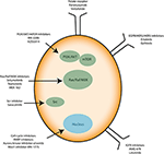
Recently, anti-angiogenic agents have been introduced into ovarian cancer treatment protocols. They target the formation of new vessels from preexisting vessels (angiogenesis), a process necessary for tumor survival and spread. Bevacizumab (Avastin®) is a monoclonal antibody that binds to the pro-angiogenic factor VEGF (Vascular Epidermal Growth Factor) and prevents its interaction with its receptors located on the surface of endothelial cells (61). In parallel, many clinical trials aim to use small inhibitory pharmacological molecules targeting the processes involved in tumor growth and/or spread (antibodies, inhibitors). Special mention of antibodies directed against pro-angiogenic factors (anti-VEGF antibodies), molecules blocking DNA repair systems (PARP inhibitor) or also inhibitors of folate receptors, the latter being overexpressed by ovarian cancer cells. Molecules that interfere with altered signaling pathways in ovarian cancer are also developed. They target the MAP-Kinases, PI3-Kinase/Akt pathways or integrin-type adhesion receptors. Immunotherapy strategies are also being considered (antibodies, inhibitors of immunological checkpoints, vaccines) (Figure 3). All these trials open up new therapeutic perspectives with the hope of seeing new molecules coming to the market for the treatment of ovarian or other type of cancer such as glioblastoma multiforme (GBM) (62).

FIGURE 3
www.frontiersin.org
Figure 3. Immunotherapies applied to ovarian cancer cells. Known therapies mainly target signaling proteins involved in cell proliferation and resistance to apoptosis such as PI3K/AKT/mTOR, Src, and Ras/Raf/MEK. Growth factor receptors and cell cycle inhibitors are used too.


Cancer Recurrence: the Special Case of GBM and Ovarian Cancer

In the case of ovarian cancer, 70–80% of patients respond well to initial treatments. However, more than 80% of patients relapse within the next 6–36 months. Second line treatments include a cytoreductive surgery combined with chemotherapy (63). Second-line treatments aim to improve the quality of life of patients and increase the progression-free survival of the disease. In the absence of effective therapies, most recurrent ovarian cancers are incurable. This is likely due to phenotypic plasticity and adaptation, a process common to cancer cells (64, 65). During the recurrence process, ovarian cancer cells acquire an adaptive phenotype allowing them to overcome the mechanisms guarantying cell and tissue homeostasis. These characteristics were identified in 2000 and supplemented in 2011 by Hanahan and Weinberg (66, 67). These characteristics are: (i) self-sufficiency in proliferative signals (ii) insensitivity to growth inhibition signals, (iii) ability to replicate indefinitely, (iv) genomic instability, (v) angiogenic induction potential, (vi) an ability to invade tissues and form metastases, (vii) a misregulation of cellular energy metabolism, (viii) an ability to escape the immune system and to promote an inflammatory environment, and (ix) the ability to resist cell death. This constitutes a major obstacle in the therapeutic management of cancers. Taken all the above, a special focus need being placed on points (I and II), and to decipher signaling pathways that could explain the phenotypic variability within a tumor mass leading to resistance to programmed cell death.

Chemoresistance of Ovarian Cancers

Chemoresistance is defined as the ability of cancer cells to survive the cytotoxic effect of anticancer agents. It can manifest itself right away, i.e., intrinsic chemoresistance. In other cases, the treatments make it possible to obtain a partial or incomplete response, which then become ineffective over time. This is defined as acquired chemoresistance. Loss of sensitivity of ovarian cancer cells to platinum-based treatments is one of the major complications of ovarian cancer. The mechanisms leading to the appearance of this chemoresistance have been classified in four categories according to the moment when they intervene in the response chain of the CDDP (68). Two mechanisms promote the inhibition of the cytotoxic effect of CDDP before its interaction with DNA. A decrease in the concentration of intracellular CDDP is observed following an inhibition of facilitated diffusion by the copper transporter or an increase in its efflux by ABC-type transporters (ATP Binding Cassette). For example, overexpression of ATP7A (a member of the ABC protein family) is associated with a poor prognosis (69). An increase in the sequestration of CDDP by thiols intracellular occurs. Indeed, glutathione (GSH) can react with CDDP thus forming a platinum-GSH complex which will be subsequently eliminated. γ-Glutamylcyteine synthetase, the enzyme responsible for the synthesis of GSH, has been shown to be overexpressed in ovarian cancers (7072).

Chemoresistance of Glioblastoma Multiforme

It is generally accepted that cancer cells produce large amounts of ROS, mainly in the inner mitochondrial membrane (IMM), the site of cellular respiration (73). When the flow of electrons is slowed down at the level of the electron transfer chain (ETC), this produces mitochondrial ROS (mtROS) species such as superoxide radical (O2.−), hydrogen peroxide (H2O2) or hydroxyl radical (.OH). These reactive species can, in the long run, lead uncontrolled oxidations of mitochondrial DNA (mtDNA), lipids and proteins. Interestingly, cancer cells have been shown to decrease the generation of mitochondrial ROS by improving their mitochondria coupling (74). This phenomenon even occurs when external ROS generation methods are applied. This is the case of glioblastoma multiforme (GBM) cells resistant to Temozolomide (TMZ), a chemotherapy drug commonly used for treating gliomas (75). Recent studies have demonstrated that the resistance of glioma cells to drugs are acquired characteristics that are linked to mitochondrial activity in general, and to the activity of complexes (I-IV) of the electron transfer chain in particular (13, 7476). Oliva et al. (75) studied chemoresistance in a glioma cell line and xenograft using TMZ chemotherapy agent. They managed to show that TMZ-resistant cells have a better management of mtROS generation due to a higher mitochondrial coupling. Moreover, the chemoresistant glioma cell line showed reduced mtROS production concomitant with increased oxygen consumption, and lowered proton leak. Interestingly, the authors showed that TMZ-resistant cells consumed less glucose and produced less lactate which are markers of reduced Warburg effect (Figure 4). Finally, the authors managed to reserve TMZ resistance and increased sensitivity to chemotherapy by treating glioma cells with the oxidant L-buthionine-S,R-sulfoximine. On the other hand, the antioxidant N-acetyl-cysteine treatment prevented TMZ cytotoxicity in sensitive cell lines by vanishing TMZ-dependent mtROS generation (75). These observations show the pivotal role of mtROS in the induction of pro and antitumor signaling pathways (74).

FIGURE 4
www.frontiersin.org
Figure 4. Mitochondrial checking of Reactive Oxygen Species (ROS) in cancer cells is crucial in tumor resistance to therapies. ROS accumulation is known to trigger cancer cell proliferation supported by nutrients consumption, manly glucose, and pronounced Warburg effect (increased lactate production). Strategic reduction of mitochondrial ROS by complex IV increases cancer cells’ robustness to external stresses such as chemotherapies.


TMZ triggers mtDNA alterations and creates local mtROS accumulation. Then, proteins and lipids are at the mercy of mtROS, which trigger its oxidation and compromise mitochondrial membrane integrity. Therefore, mtROS seems to have a feed-forward loop effect on mtDNA. Indeed, it has been reported that minor mtDNA alterations and mutations are major contributors to mtROS accumulation in cancer cells (77). Exactly 13 ETC proteins are encoded in mtDNA and the rest are nuclear genome encoded (78). Mutations in the genes encoding these subunits cause ETC dysfunction and accumulation of mtROS (74, 79). Interestingly, Oliva et al. (75) also reported a sharp drop of TMZ-induced ROS generation in mitochondrial DNA-depleted (ρ°) glioma cell line and decreased cell sensitivity to TMZ drug. Sensitivity of the glioma cell line to TMZ was restored upon repopulating cell with functioning mitochondria. As we postulated above, mitochondrial metabolism and its integrity appear to play a pivotal role in the treatment of cancer and recurrences that may occur.


Metabolic Therapies for Cancer Treatment

One of the most studied pathology related to mitochondrial metabolism is cancer. First studies date from the 1920s in which Otto Heinrich Warburg, future Nobel Prize of medicine winner in 1931, observes that cancer cells produce abnormally high amounts of lactate, even in oxygen aerated environment (9, 71, 80). He explained that cancer cells derive their energy mainly from the fermentation of glucose or aerobic glycolysis. Warburg later hypothesized that the fermentative phenotype of cancer cells was due to dysfunction of the mitochondria (8). Our recent works has helped into validation of some Warburg’s assumptions. Indeed, we have demonstrated that human and mouse cancer cell lines have very low mitochondrial membrane potential (ΔΨm) and more pronounced glycolysis compared to the respective healthy cells (81). We also proposed complementary therapeutic approaches to chemotherapy, aiming to counteract the Warburg effect in cancer cells (34, 8284). These are METABLOC, a combination of small molecules composed of α-lipoic acid, pushing carbon flux to mitochondria, and hydroxycitrate, an inhibitor of lipogenesis.

Cancer Cells Have a Reduced Mitochondria Horsepower

Cancer cell growth is promoted by the anabolic signaling pathways and sustained metabolic reprogramming (45, 85, 86). We sought to establish the metabolic profile of cancer cells based on the characterization of physico-chemical parameters in healthy and cancerous primary cells isolated from the colon in patients. For each of the two populations, cells were separated by elutriation and then collected in different phases of the cell cycle (G0/G1/S/G2/M) according to their sizes. The parameters studied are the redox potential of the cells by quantification of the NA(D)/NAD(P)H species, the energetic state of the cells by ATP assay and the intracellular pH (pHi), which could be associated with the metabolic activity. The results confirm the two major phenomena associated with cancer cells: the metabolic activity is more pronounced compared to healthy cells, which results in a more alkaline pHi (81). Indeed, cancer populations in G0 have, on average, a pHi of 7.29 ± 0.13 while the pHi of healthy cells in the same phase of the cycle is 6.87 ± 0.10. We have also reported a lower energy efficiency in the cancer population. This is characterized by a lower amount of ATP (2X less in G0). The NAD+/NADH and NADP+/NADPH redox ratios are up to 5X and 10X higher, respectively, in cancer cells. It also reflects a more pronounced glycolytic flow as hypothesized by Warburg, while the production of lactate in the culture medium has not been determined. Indeed, a higher NAD+/NADH ratio is necessary for the maintenance of glycolysis while the production of lactate ensures the turnover of NAD+ by lactate dehydrogenase (LDH). Likewise, NADP+/NADPH conditions entry into the pentose phosphate pathway (PPP) and generates the NADPH necessary for the synthesis of fatty acids and of other membrane lipids. These results were then confirmed in various mice and human cell lines. In addition, ΔΨm has been quantified in healthy and cancerous cell lines. The results show that the mitochondrial membrane potential is significantly lower in cancer lines. This lower ΔΨm in cancer cells is assumed, such as lower energy efficiency (ATP synthesis), even when higher glycolytic flux is found in cancer cell population. Therefore, these results partially confirm Warburg’s observations and offer avenues for therapeutic innovations.

Metabolic Therapies Targeting Tumor Growth

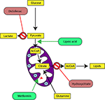
In our last experimental study, we followed tumor growth in mice to which tumors were grafted and subjected to metabolic treatments for 59 days (84). The molecules used part of the METABLOC, supplemented with other molecules known from the pharmacopeia. These are α-lipoic acid (α-LA) and hydroxycitrate (HCA), both used as food supplements. The α-LA is an inhibitor of pyruvate dehydrogenase kinase-2 (PDK2), reported as inhibitor of pyruvate dehydrogenase (PDH) in normal cells but to a greater extent in cancer cell (85, 87, 88) (Figure 5). Interestingly, a recent study also reported effective inhibition of angiogenesis and HIF1-α activity in mice tumor xenograft under dichloroacetate (DCA) treatment (89), known as a major inhibitor of PDK2 (90). Moreover, PDK2 gene disruption in lung cancer cells have been reported to increase cell sensitivity to Paclitaxel chemotherapeutic agent (91). Part of METABLOC, HCA is an inhibitor of ATP-citrate lyase (ACL) to prevent lipogenesis (82, 92, 93). Finally, we used metformin, which is used in type II diabetes (94) and reported as inhibitor of complex I (9597), and diclofenac, which is an anti-inflammatory and inhibitor of lactate dehydrogenase (LDH) and of the transporter of monocarboxylate (MCT1) (98, 99). A positive control group of mice was treated with cisplatin, a classic chemotherapeutic agent. We reported that the combination of these four molecules has an inhibitory effect on the growth of the tumor implanted in mice (84). Indeed, when the molecules are used separately, there are no major effects compared to the control group without treatment. However, when metformin is applied in high doses combined with α-LA and HCA, the growth of the tumor is clearly slowed down and then inhibited after ~50 days of follow-up. The inhibition is even more pronounced when high-dose diclofenac is added in combination with METABLOC and high-dose metformin.

FIGURE 5
 
www.frontiersin.org
Figure 5. Cancer metabolic therapy targets central carbon metabolism and enhances mitochondrial activity. Diclofenac is an inhibitor of the Lactate dehydrogenase (LDH). By inhibition of dehydrogenase kinase-2 (PDK2), Lipoic acid promotes Pyruvate dehydrogenase (PDH) activity. Metformin activates mitochondrial uncoupling proteins and triggers mitochondrial membrane depolarization. Consequently, it increases electron transfer chain activities. Hydroxycitrate is an inhibitor of ATP-citrate lyase (ACL) and prevents cytosolic Acetyl-CoA (AcCoA) accumulation as precursor for lipogenesis.
 

In parallel with this experimental work, we have developed a kinetic model of central metabolism to simulate tumor growth and predict the effect of previous molecules on intracellular energy flows. This model includes a reduced and simplified metabolic network of cancer cell metabolism and manages to simulate tumor growth but also the inhibitory effect of the metabolic therapy. Simulations show a reverse Warburg effect under the action of metabolic therapy. This is evidenced by a net flow of blood lactate re-consumption through LDH and increased mitochondrial respiration, a characteristic phenotype of healthy cells. These experimental results show that it is possible to perturb the metabolic stability of cancer cells and restore a basal metabolism close to that of healthy cells. Mitochondrial metabolism in general, and cell respiration through the electron transfer chain in particular, therefore seems to be the ideal target to make tumor masses more vulnerable to treatments and for inducing apoptosis or necrosis of the tumor mass (84).


Perspective: Over-Activating Cancer Cells Mitochondria by Singlet Oxygen-Oriented Photodynamic Therapy

As summarized in the previous sections, the synthesis of ATP by OXPHOS is made possible thanks to the successive transfers of electrons through the electron transfer chain (ETC) (Figure 1). Misregulation on one of these ETC complexes often causes metabolic disorders ranging from transient paralysis to prolonged degenerative processes such as myopathies, Alzheimer’s, cancer, and other acquired degenerations (41). In the case of cancer, we have reported works showing the strict regulations of these complexes as well as the strategies adopted by cancer cells to escape from the immune system regulation. We bring here a new explanation, under strong assumptions, on the “mitochondrial dysfunctions” observed in certain sub-populations of cells as well as therapeutic solutions which could reverse the degenerative process linked to the proliferation and metastasis of most of tumor cells.

We propose that saturation of ETC with electrons could be the cause of the glycolytic phenotype of cancer cells but also of the accumulation of deleterious oxidizing species, such as ROS, also present in neurodegenerative pathologies (Alzheimer, Parkinson, Huntington). This electrochemical engorgement of the respiratory chain can as well be explained by the inhibition of complexes of the ETC which would prevent the transit of the electrons and by a modification of the physicochemical nature of oxygen (the final acceptor of the electrons in the ETC). Thus, cancer cells carry out a metabolic reprogramming to reach out a new metabolic steady state characterized by rapid proliferation and pronounced glycolysis, a specific phenotype described by DeBerardinis and Chandel (7) and Warburg (8). Our undisclosed preliminary results support the hypothesis of a modification of the energetic state of intracellular oxygen to explain an overvoltage at the “limits” of ETC.

Oxygen Activation in Biological Systems

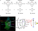
Despites its high thermodynamic reactivity, dioxygen reacts slowly with most organic molecules because of spin restriction. Its stable state corresponds to a triplet electronic ground state, referred as triplet oxygen (3O2). The stable 3O2 has two unpaired electrons occupying the π molecular orbitals with the same spin orientation (Figure 6A). This prevents its spontaneous combustion with molecules having paired electrons (100). On the other hand, O2 can turn highly reactive with an input of energy and electronic excitation of the ground state 3O2. This excited state is referred as singlet oxygen (1O2) in which the electrons of the π orbitals are paired with opposite spin. This allows 1O2 to be much more reactive with organic molecules (101) (Figure 6A).

FIGURE 6
 
www.frontiersin.org
Figure 6. Proposed model of dioxygen reduction by cytochrome c oxidase (CcO) and potential role of combined light-methylene blue (MB)-induced singlet oxygen (1O2) generation. (A) Triplet and singlet oxygen molecular orbitals representation. Triplet oxygen (3∑) is the ground state, with unpaired spins at π* orbital. Singlet oxygens (1Δ and 1∑) are the excited state with opposite spins at π* orbital. (B) The respiratory dioxygen reduction by CcO is ensured by the binuclear center with Heme a3 and CuB. The catalytic cycle of O2 reduction to water molecules (H2O) is shown from R to EH state. This cyclic model is proposed by Ishigami et al. (23). We obtained the kind permission of the corresponding author to use part of their figure initially in their study. In brief, the full cycle uses one O2 molecule, four electrons, four protons, and allows the pumping of additional four protons from the mitochondrial matrix to the intermembrane space. Two H2O molecules are produced. Electrons are sequentially transferred from the ETC cytochrome c CuA Heme a. Dioxygen first binds to heme a3 iron atom in reduced state (R) to form an intermediate state (A). Then one electron is transferred from heme a to heme a3-iron and another one form CuB for the cleavage of O-O bonds. Follows sequential electrons and protons transfer until complete O2 reduction to water molecules. For more detailed description, see (23). We proposed that during the oxidative phase the oxygen molecules assume different oxidative forms with the iron atom which promote the production of ROS species (1O2,.OH,.O.2) during disturbances in the mitochondrial membrane due to toxic substances or instability of the lipid bilayer. Although the exact mechanism of formation of these ROS remains hypothetical, we propose a phototherapeutic approach consisting in the controlled generation of singlet oxygen via methylene blue (MB) or other photosensitizers. In this illustrating example, MB is in triplet (3MB) state in natural condition. When exposed to laser irradiation at specific wavelength (630–680 nm, <5 mW), 3MB reaches its excited 1MB state and triggers 1O2 generation. 1MB or other photosensitizers could be used for CcO-mimiking 3O2 activation and 1O2 generation in cancer cells. Mitochondrial singlet oxygen accumulation may rescue the Warburg phenotype and trigger cancer cell death.
 

Generation of 1O2 from water molecule have been widely reported during photosynthesis in plants and cyanobacteria, using energy from the sunlight (102). In these systems, singlet oxygen is produced by light absorption by the photosensitizers. It is especially the case in plants where 1O2 is generated by the chlorophylls and other cofactors of the photosystem II (103). Once the highly reactive 1O2 is produced, it can be deactivated by quenching molecules such as beta-carotene, alpha-tocopherol, or plastoquinone. Cytochrome complex has also been involved in 1O2 production in plant (104). The authors showed that photoactivation of isolated cytochrome b6f complex triggers 1O2 generation. More precisely, it was shown the Rieske Fe-S protein-like cytochrome b6f center is the cluster involved in 1O2 production (31). Taken together, these observations raise the question of a possible involvement of singlet oxygen in mitochondrial respiration.

The eukaryotic respiratory chain has been extensively studied. As depicted in previous sections, it is essentially composed of five protein complexes involved in electron transfer, proton pumping across the mitochondrial membrane, oxygen reduction to water by the complex IV or cytochrome c oxidase (CcO), and ATP synthesis by ATP synthase (17, 25, 105108). Three parameters have been proposed as key in controlling cell respiration: mitochondrial membrane pH gradient (ΔpH), O2 concentration and [ferricytochrome c]/[ferrocytochrome c] ratio at the ETC (109). Arnold et al. (109) first reported that CcO subunits stability is mediated by cardiolipin and essential for the regulations. They found that high mitochondrial matrix ATP-to-ADP ratio has an allosteric feedback inhibition on the complex IV and cell respiration. However, partial or total inhibition of the respiratory chain is one of the main causes of production of mitochondrial reactive oxygen species (ROS) (73, 74) and of actively-promoting mitochondrial metabolic switch supporting tumor progression and metastasis (13). Interestingly, singlet oxygen is reported as the main ROS produced during OXPHOS in yeast and human healthy and cancer cells’ mitochondria (110). Its high reactive potential causes protein and mitochondrial DNA (mtDNA) damages (111, 112). However, new findings support the idea of metabolic and signaling activities of a mitochondrial low dose of 1O2 (113). Zhou et al. (113) triggered 1O2 generation in HeLa cancer cell line by laser irradiation and showed increased mtDNA replication, which is also a marker of increased mitochondrial respiration. Interestingly enough, pioneering studies showed that fast electron transfer through eukaryotes and prokaryotes electron transfer chain (ETC) achieves high O2 affinity to CcO (19, 21). More recently, studies have depicted the most probable mechanism beyond O2 activation by CcO complex (24, 37). CcO ensures cell respiration by sustaining both protons translocation and O2 reduction to water molecules. O2 activation is catalyzed by the binuclear heme-copper active site in a catalytic cycle by addition of four electrons routed through ETC (Figure 6B). Full O2 reduction is coupled with four protons translocation though the inner mitochondrial membrane (IMM) as showed in Figure 1 (20). The complete O2 reduction cycle model has been well depicted by (23), and the mitochondrial membrane stability seems to be a key parameter in oxygen activation and ROS generation (114).

Singlet Oxygen-Oriented Photodynamic Therapy

Taking all the above, we here propose the development of a novel therapy targeting singlet oxygen using photodynamic techniques. The expected outcome is 1O2 or other ROS-induced cancer cells apoptosis and tumor regression as nicely reported in recent studies by the mean of extracellular singlet oxygen generation (115, 116). To do so, we propose that photosensitizers such as methylene blue (MB), chlorophyll, and protoporphyrin could play an intermediary role in the electron decongestion of ETC by catalyzing the activation of 3O2 into 1O2 and thus promoting apoptosis by accumulation of ROS species (Figure 6B). All these photosensitizers could be highly excited with a light source at specific wave lengths. MB has a double application. It is used as a dye in the textile industry, as well as a medicine for its antimicrobial properties and also applied as an antidote during cyanide, inhibitor of the complex IV (CIV), poisoning, or in cases of methemoglobinemia (117). Therefore, when administered MB acts as a CIV in the reduction of dioxygen to the water molecules (118) (Figure 6B). This singlet oxygen-oriented photodynamic therapy (PDT) is thought as a mimicry to chemotherapies such as does animals with predators (113, 119, 120). In addition, we propose that 1O2-oriented PDT could increase sensitivity of tumor cells, specially the resistant and ones, to conventional therapies. Furthermore, our recent study in Chinese Hamster Ovary (CHO) cells supports this hypothesis. Indeed, we have reported that combination of MB and METABLOC reduced the Warburg effect in CHO and optimized monoclonal antibody (mAB) production, which is a marker of an increased mitochondrial OXPHOS (121). Finally, we strongly believe there are experimental evidences that the resistance of tumor to conventional treatments may be overcome by targeting cancer cells’ “Achilles’ heel,” the ROS accumulation, by introducing photosensitizers as “trojan horses.”


Conclusion

Mitochondria are much more than just a factory for producing energy for the cell. It is a cornerstone between the three essential processes for maintaining the stability of a multicellular organism: proliferation, differentiation, and cell death. Poor regulation of asymmetric cell proliferation often leads to local destabilization of tissues and degenerates into tumor masses that escape all regulations by the immune system. Therapeutic approaches such as chemotherapies, metabolic therapies, immunotherapies, or even radiotherapy make it possible to eliminate the most fragile cancer cells. However, mitochondria confer the cells a great capacity for adaptation and resistance to these agents perceived as external stresses. Our proposals are a call to develop soft skills methods to hijack cancer cell metabolism. This involves the use of molecules easily ingested by cancer cells and perceived as beneficial to meet their energy demand. This is the case with photosensitizers like methylene blue, protoporphyrin or chlorophyll already implemented in plants. In the second step, one will be applying the photodynamic therapy in order to excite these molecules present in the cell. They will create highly reactive species of which singlet oxygen (1O2) seems to be the main produced at the level of the CI, CIII, and perhaps CIV complexes of the ETC. We speculate that the accumulation of 1O2 in mitochondria will trigger apoptosis in cancer cells, especially those that are resistant to conventional treatments.


Author Contributions

JV wrote the manuscript and draw the figures. LS and MJ contributed to the thinking and reviewed the manuscript. All authors contributed to the article and approved the submitted version.


Funding

This work has been supported by the French association Guérir du cancer empowered by Fondation de France and the Fondation et Alumni de Polytechnique Montréal.


Conflict of Interest

The authors declare that the research was conducted in the absence of any commercial or financial relationships that could be construed as a potential conflict of interest.


Acknowledgments

The authors thank Dr. Lyvia Blay for interesting discussions on chemoresistance.


References

1. Lane N. Oxygen: The Molecule That Made the World. (2003) Oxford, UK: Oxford University Press.
Google Scholar


2. Blázquez-Castro A. Direct 1O2 optical excitation: a tool for redox biology. Redox Biol. (2017) 13:39–59. doi: 10.1016/j.redox.2017.05.011
PubMed Abstract | CrossRef Full Text | Google Schola


3. Gonzalez H, Hagerling C, Werb Z. Roles of the immune system in cancer: from tumor initiation to metastatic progression. Genes Develop. (2018) 32:1267–84. doi: 10.1101/gad.314617.118
PubMed Abstract | CrossRef Full Text | Google Scholar


4. Keenan M, Chi J-T. Alternative fuels for cancer cells. Cancer J. (2015) 21:49–55. doi: 10.1097/PPO.0000000000000104
PubMed Abstract | CrossRef Full Text | Google Scholar


5. von Neumann J, Morgenstern O. Theory of Games and Economic Behavior. Princeton: Princeton University Press. (1944).
Google Scholar


6. Smith JM, Maynard Smith J, Holliday R. Game theory and the evolution of behaviour. Proc R Soc Lond Series B Biol Sci. (1979) 205:475–88. doi: 10.1098/rspb.1979.0080
PubMed Abstract | CrossRef Full Text | Google Scholar


7. DeBerardinis RJ, Chandel NS. We need to talk about the Warburg effect. Nat Metab. (2020) 2:127–9. doi: 10.1038/s42255-020-0172-2
PubMed Abstract | CrossRef Full Text | Google Scholar


8. Warburg O. On the origin of cancer cells. Science. (1956) 123:309–14. doi: 10.1126/science.123.3191.309
PubMed Abstract | CrossRef Full Text | Google Scholar


9. Warburg O. The metabolism of carcinoma cells. J Cancer Res. (1925) 9:148–63. doi: 10.1158/jcr.1925.148
CrossRef Full Text | Google Scholar


10. Cassim S, Vučetić M, Ždralević M, Pouyssegur J. Warburg and beyond: the power of mitochondrial metabolism to collaborate or replace fermentative glycolysis in cancer. Cancers. (2020) 12:1119. doi: 10.3390/cancers12051119
CrossRef Full Text | Google Scholar


11. Weinberg F, Chandel NS. Mitochondrial metabolism and cancer. Ann N Y Acad Sci. (2009) 1177:66–73. doi: 10.1111/j.1749-6632.2009.05039.x

PubMed Abstract | CrossRef Full Text | Google Scholar


12. Guerra F, Arbini AA, Moro L. Mitochondria and cancer chemoresistance. Biochim Et Biophys Acta Bioenerg. (2017) 1858:686–99. doi: 10.1016/j.bbabio.2017.01.012
PubMed Abstract | CrossRef Full Text | Google Scholar


13. Porporato PE, Payen VL, Pérez-Escuredo J, De Saedeleer CJ, Danhier P, Copetti T, et al. A mitochondrial switch promotes tumor metastasis. Cell Rep. (2014) 8:754–66. doi: 10.1016/j.celrep.2014.06.043
PubMed Abstract | CrossRef Full Text | Google Scholar


14. da Veiga Moreira J, Peres S, Steyaert J-M, Bigan E, Paulevé L, Nogueira ML, et al. Cell cycle progression is regulated by intertwined redox oscillators. Theor Biol Med Model. (2015) 12:10. doi: 10.1186/s12976-015-0005-2
PubMed Abstract | CrossRef Full Text | Google Scholar


15. Mitra K, Wunder C, Roysam B, Lin G, Lippincott-Schwartz J. A hyperfused mitochondrial state achieved at G1–S regulates cyclin E buildup and entry into S phase. Proc Natl Acad Sci USA. (2009) 106:11960–5. doi: 10.1073/pnas.0904875106
PubMed Abstract | CrossRef Full Text | Google Scholar


16. Weinberg SE, Sena LA, Chandel NS. Mitochondria in the regulation of innate and adaptive immunity. Immunity. (2015) 42:406–17. doi: 10.1016/j.immuni.2015.02.002
PubMed Abstract | CrossRef Full Text | Google Scholar


17. Mitchell P. Chemiosmotic coupling in oxidative and photosynthetic phosphorylation. Biol Rev. (1961) 41:445–501. doi: 10.1111/j.1469-185X.1966.tb01501.x
PubMed Abstract | CrossRef Full Text | Google Scholar


18. Schultz BE, Chan SI. Structures and proton-pumping strategies of mitochondrial respiratory enzymes. Annu Rev Biophys Biomol Struct. (2001) 30:23–65. doi: 10.1146/annurev.biophys.30.1.23

PubMed Abstract | CrossRef Full Text | Google Scholar


19. Verkhovsky MI, Morgan JE, Puustinen A, Wikström M. Kinetic trapping of oxygen in cell respiration. Nature. (1996) 380:268–70. doi: 10.1038/380268a0
PubMed Abstract | CrossRef Full Text | Google Scholar


20. Wikström M, Sharma V. Proton pumping by cytochrome c oxidase – a 40 year anniversary. Biochim et Biophys Acta (BBA) – Bioenerg. (2018) 1859:692–8. doi: 10.1016/j.bbabio.2018.03.009
PubMed Abstract | CrossRef Full Text | Google Scholar


21. Wikstrom MKF. Proton pump coupled to cytochrome c oxidase in mitochondria. Nature. (1977) 266:271–3. doi: 10.1038/266271a0
PubMed Abstract | CrossRef Full Text | Google Scholar


22. Tomashek JJ, Brusilow WS. Stoichiometry of energy coupling by proton-translocating ATPases: a history of variability. J Bioenerg Biomembr. (2000) 32:493–500. doi: 10.1023/A:1005617024904
PubMed Abstract | CrossRef Full Text | Google Scholar


23. Ishigami I, Lewis-Ballester A, Echelmeier A, Brehm G, Zatsepin NA, Grant TD, et al. Snapshot of an oxygen intermediate in the catalytic reaction of cytochrome c oxidase. Proc Natl Acad Sci USA. (2019) 116:3572–7. doi: 10.1073/pnas.1814526116
PubMed Abstract | CrossRef Full Text | Google Scholar


24. Wikström M, Krab K, Sharma V. Oxygen activation and energy conservation by Cytochrome c oxidase. Chem Rev. (2018) 118:2469–90. doi: 10.1021/acs.chemrev.7b00664

PubMed Abstract | CrossRef Full Text | Google Scholar


25. Mazat J-P, Ransac S, Heiske M, Devin A, Rigoulet M. Mitochondrial energetic metabolism—some general principles. IUBMB Life. (2013) 65:171–9. doi: 10.1002/iub.1138
PubMed Abstract | CrossRef Full Text | Google Scholar


26. Heinrich R, Rapoport TA. A linear steady-state treatment of enzymatic chains. general properties, control and effector strength. Eur J Biochem. (1974) 42:89–95. doi: 10.1111/j.1432-1033.1974.tb03318.x
PubMed Abstract | CrossRef Full Text | Google Scholar


27. Kacser H, Burns JA. The control of flux. Biochem Soc Trans. (1995) 23:341–66. doi: 10.1042/bst0230341
PubMed Abstract | CrossRef Full Text | Google Scholar


28. Reder C. Metabolic control theory: a structural approach. J Theor Biol. (1988) 135:175–201. doi: 10.1016/S0022-5193(88)80073-0
PubMed Abstract | CrossRef Full Text | Google Scholar


29. Bohnensack R, Küster U, Letko G. Rate-controlling steps of oxidative phosphorylation in rat liver mitochondria. a synoptic approach of model and experiment. Biochim Et Biophys Acta. (1982) 680:271–80. doi: 10.1016/0005-2728(82)90139-6
PubMed Abstract | CrossRef Full Text | Google Scholar


30. Kholodenko BN. Control of mitochondrial oxidative phosphorylation. J Theor Biol. (1984) 107:179–88. doi: 10.1016/S0022-5193(84)80020-X
PubMed Abstract | CrossRef Full Text | Google Scholar


31. Rieske JS, MacLennan DH, Coleman R. Isolation and properties of an iron-protein from the (reduced coenzyme Q)-cytochrome C reductase complex of the respiratory chain. Biochem Biophys Res Commun. (1964) 15:338–44. doi: 10.1016/0006-291X(64)90171-8
CrossRef Full Text | Google Scholar


32. Cecchini G. Function and structure of Complex II of the respiratory chain. Annu Rev Biochem. (2003) 72:77–109. doi: 10.1146/annurev.biochem.72.121801.161700
PubMed Abstract | CrossRef Full Text | Google Scholar


33. Sazanov LA, Hinchliffe P. Structure of the hydrophilic domain of respiratory complex I from thermus thermophilus. Science. (2006) 311:1430–6. doi: 10.1126/science.1123809
PubMed Abstract | CrossRef Full Text | Google Scholar


34. Lauble H, Kennedy MC, Beinert H, Stout CD. Crystal structures of aconitase with isocitrate and nitroisocitrate bound. Biochemistry. (1992) 31:2735–48. doi: 10.1021/bi00125a014
PubMed Abstract | CrossRef Full Text | Google Scholar


35. Graham LA, Trumpower BL. Mutational analysis of the mitochondrial Rieske iron-sulfur protein of Saccharomyces cerevisiae. III. import, protease processing, and assembly into the cytochrome bc1 complex of iron-sulfur protein lacking the iron-sulfur cluster. J Biol Chem. (1991) 266:22485–92.
PubMed Abstract | Google Scholar


36. Diaz F, Kotarsky H, Fellman V, Moraes CT. Mitochondrial disorders caused by mutations in respiratory chain assembly factors. Semin Fetal Neonatal Med. (2011) 16:197–204. doi: 10.1016/j.siny.2011.05.004
PubMed Abstract | CrossRef Full Text | Google Scholar


37. Wikström M, Sharma V. Oxygen reduction and proton translocation by respiratory Cytochrome c Oxidase. In: M. Ikeda-Saito, E. Raven, editors. Dioxygen-Dependent Heme Enzymes. Cambridge: Royal Society of Chemistry (2018). p. 311–33. doi: 10.1039/9781788012911-00311
CrossRef Full Text | Google Scholar


38. Schäfer E, Seelert H, Reifschneider NH, Krause F, Dencher NA, Vonck J. Architecture of active mammalian respiratory chain supercomplexes. J Biol Chem. (2006) 281:15370–5. doi: 10.1074/jbc.M513525200
PubMed Abstract | CrossRef Full Text | Google Scholar


39. Chen D, Roda JM, Marsh CB, Eubank TD, Friedman A. Hypoxia inducible factors-mediated inhibition of cancer by GM-CSF: a mathematical model. Bull Math Biol. (2012) 74:2752–77. doi: 10.1007/s11538-012-9776-3

PubMed Abstract | CrossRef Full Text | Google Scholar


40. Strogolova V, Furness A, Robb-McGrath M, Garlich J, Stuart RA. Rcf1 and Rcf2, members of the hypoxia-induced Gene 1 protein family, are critical components of the mitochondrial Cytochrome bc1-Cytochrome c oxidase supercomplex. Mol Cell Biol. (2012) 32:1363–73. doi: 10.1128/MCB.06369-11
PubMed Abstract | CrossRef Full Text | Google Scholar


41. Lenaz G, Genova ML. Supramolecular organisation of the mitochondrial respiratory chain: a new challenge for the mechanism and control of oxidative phosphorylation. In: Kadenbach B, editor. Mitochondrial Oxidative Phosphorylation: Nuclear-Encoded Genes, Enzyme Regulation, and Pathophysiology. New York, NY: Springer (2012). p. 107–44. doi: 10.1007/978-1-4614-3573-0_5
CrossRef Full Text | Google Scholar


42. Vartak R, Porras CA-M, Bai Y. Respiratory supercomplexes: structure, function and assembly. Protein Cell. (2013) 4:582–90. doi: 10.1007/s13238-013-3032-y
PubMed Abstract | CrossRef Full Text | Google Scholar


43. DeBerardinis RJ, Lum JJ, Hatzivassiliou G, Thompson CB. The biology of cancer: metabolic reprogramming fuels cell growth and proliferation. Cell Metab. (2008) 7:11–20. doi: 10.1016/j.cmet.2007.10.002
PubMed Abstract | CrossRef Full Text | Google Scholar


44. DeBerardinis RJ, Chandel NS. Fundamentals of cancer metabolism. Sci Adv. (2016) 2:e1600200. doi: 10.1126/sciadv.1600200
PubMed Abstract | CrossRef Full Text | Google Scholar


45. Faubert B, Solmonson A, DeBerardinis RJ. Metabolic reprogramming and cancer progression. Science. (2020) 368:eaaw5473. doi: 10.1126/science.aaw5473
PubMed Abstract | CrossRef Full Text | Google Scholar


46. Lapuente-Brun E, Moreno-Loshuertos R, Acín-Pérez R, Latorre-Pellicer A, Colás C, Balsa E, et al. Supercomplex assembly determines electron flux in the mitochondrial electron transport chain. Science. (2013) 340:1567–70. doi: 10.1126/science.1230381
PubMed Abstract | CrossRef Full Text | Google Scholar


47. McGuire WP, Hoskins WJ, Brady MF, Kucera PR, Partridge EE, Look KY, et al. Cyclophosphamide and cisplatin compared with paclitaxel and cisplatin in patients with Stage III and Stage IV ovarian cancer. N Engl J Med. (1996) 334:1–6. doi: 10.1056/NEJM199601043340101
PubMed Abstract | CrossRef Full Text | Google Scholar


48. Piccart MJ, Green JA, Lacave AJ, Reed N, Vergote I, Benedetti-Panici P, et al. Oxaliplatin or paclitaxel in patients with platinum-pretreated advanced ovarian cancer: a randomized Phase II study of the european organization for research and treatment of cancer gynecology group. J Clin Oncol. (2000) 18:1193–202. doi: 10.1200/JCO.2000.18.6.1193
CrossRef Full Text | Google Scholar


49. Rosenberg B, Camp LV, Krigas T. Inhibition of cell division in Escherichia coli by electrolysis products from a platinum electrode. Nature. (1965) 205:698–9. doi: 10.1038/205698a0
PubMed Abstract | CrossRef Full Text | Google Scholar


50. Rosenberg B, Vancamp L, Trosko JE, Mansour VH. Platinum compounds: a new class of potent antitumour agents. Nature. (1969) 222:385–6. doi: 10.1038/222385a0
PubMed Abstract | CrossRef Full Text | Google Scholar


51. Rosenberg B, VanCamp L. The successful regression of large solid sarcoma 180 tumors by platinum compounds. Cancer Res. (1970) 30:1799–802.
PubMed Abstract | Google Scholar


52. Higby DJ, Higby DJ, Wallace HJ, Albert DJ, Holland JF. Diaminodichloroplatinum: a phase I study showing responses in testicular and other tumors. Cancer. (1974) 33:1219–25. doi: 10.1002/1097-0142(197405)33:5&lt;1219::AID-CNCR2820330505&gt;3.0.CO;2-U
PubMed Abstract | CrossRef Full Text | Google Scholar


53. Ishida S, Lee J, Thiele DJ, Herskowitz I. Uptake of the anticancer drug cisplatin mediated by the copper transporter Ctr1 in yeast and mammals. Proc Natl Acad Sci USA. (2002) 99:14298–302. doi: 10.1073/pnas.162491399
PubMed Abstract | CrossRef Full Text | Google Scholar


54. Martinho N, Santos TCB, Florindo HF, Silva LC. Cisplatin-membrane interactions and their influence on platinum complexes activity and toxicity. Front Physiol. (2018) 9:1898. doi: 10.3389/fphys.2018.01898
PubMed Abstract | CrossRef Full Text | Google Scholar


55. Fuertes MA, Castilla J, Alonso C, Pérez JM. Cisplatin biochemical mechanism of action: from cytotoxicity to induction of cell death through interconnections between apoptotic and necrotic pathways. Curr Med Chem. (2003) 10:257–66. doi: 10.2174/0929867033368484
PubMed Abstract | CrossRef Full Text | Google Scholar


56. Stewart AL, Teno J, Patrick DL, Lynn J. The concept of quality of life of dying persons in the context of health care. J Pain Symptom Manage. (1999) 17:93–108. doi: 10.1016/S0885-3924(98)00131-6
PubMed Abstract | CrossRef Full Text | Google Scholar


57. Bishop JF, Dewar J, Toner GC, Smith J, Tattersall MH, Olver IN, et al. Initial paclitaxel improves outcome compared with CMFP combination chemotherapy as front-line therapy in untreated metastatic breast cancer. J Clin Oncol. (1999) 17:2355–64. doi: 10.1200/JCO.1999.17.8.2355
PubMed Abstract | CrossRef Full Text | Google Scholar


58. Jordan MA, Wilson L. Microtubules as a target for anticancer drugs. Nat Rev Cancer. (2004) 4:253–65. doi: 10.1038/nrc1317
PubMed Abstract | CrossRef Full Text | Google Scholar


59. Orr GA, Verdier-Pinard P, McDaid H, Horwitz SB. Mechanisms of taxol resistance related to microtubules. Oncogene. (2003) 22:7280–95. doi: 10.1038/sj.onc.1206934
PubMed Abstract | CrossRef Full Text | Google Scholar


60. Rao S, Krauss NE, Heerding JM, Swindell CS, Ringel I, Orr GA, et al. 3′-(p-azidobenzamido)taxol photolabels the N-terminal 31 amino acids of beta-tubulin. J Biol Chem. (1994) 269:3132–4.
PubMed Abstract | Google Scholar


61. Banerjee S, Kaye SB. New strategies in the treatment of ovarian cancer: current clinical perspectives and future potential. Clin Cancer Res. (2013) 19:961–8. doi: 10.1158/1078-0432.CCR-12-2243
PubMed Abstract | CrossRef Full Text | Google Scholar


62. Boussiotis VA, Charest A. Immunotherapies for malignant glioma. Oncogene. (2018) 37:1121–41. doi: 10.1038/s41388-017-0024-z
PubMed Abstract | CrossRef Full Text | Google Scholar


63. Davis A, Tinker AV, Friedlander M. “Platinum resistant” ovarian cancer: what is it, who to treat and how to measure benefit? Gynecol Oncol. (2014) 133:624–31. doi: 10.1016/j.ygyno.2014.02.038
PubMed Abstract | CrossRef Full Text | Google Scholar


64. Emmons MF, Faião-Flores F, Smalley KSM. The role of phenotypic plasticity in the escape of cancer cells from targeted therapy. Biochem Pharmacol. (2016) 122:1–9. doi: 10.1016/j.bcp.2016.06.014
PubMed Abstract | CrossRef Full Text | Google Scholar


65. Lorenzi T, Chisholm RH, Clairambault J. Tracking the evolution of cancer cell populations through the mathematical lens of phenotype-structured equations. Biol Direct. (2016) 11:43. doi: 10.1186/s13062-016-0143-4
PubMed Abstract | CrossRef Full Text | Google Scholar


66. Hanahan D, Weinberg RA. The hallmarks of cancer. Cell. (2000) 100:57–70. doi: 10.1016/S0092-8674(00)81683-9
PubMed Abstract | CrossRef Full Text | Google Scholar


67. Hanahan D, Weinberg RA. Hallmarks of cancer: the next generation. Cell. (2011) 144:646–74. doi: 10.1016/j.cell.2011.02.013
PubMed Abstract | CrossRef Full Text | Google Scholar


68. Galluzzi L, Vacchelli E, Pedro J-MB-S, Buqué A, Senovilla L, Baracco EE, et al. Classification of current anticancer immunotherapies. Oncotarget. (2014) 5:12472–508. doi: 10.18632/oncotarget.2998
PubMed Abstract | CrossRef Full Text | Google Scholar


69. Samimi G, Varki NM, Wilczynski S, Safaei R, Alberts DS, Howell SB. Increase in expression of the copper transporter ATP7A during platinum drug-based treatment is associated with poor survival in ovarian cancer patients. Clin Cancer Res. (2003) 9:5853–9.
PubMed Abstract | Google Scholar


70. Bansal A, Simon MC. Glutathione metabolism in cancer progression and treatment resistance. J Cell Biol. (2018) 217:2291–8. doi: 10.1083/jcb.201804161
PubMed Abstract | CrossRef Full Text | Google Scholar


71. Hanigan MH. Gamma-glutamyl transpeptidase: redox regulation and drug resistance. Adv Cancer Res. (2014) 122:103–41. doi: 10.1016/B978-0-12-420117-0.00003-7
PubMed Abstract | CrossRef Full Text | Google Scholar


72. Nunes SC, Serpa J. Glutathione in ovarian cancer: a double-edged sword. Int J Mol Sci. (2018) 19:E1882. doi: 10.3390/ijms19071882
PubMed Abstract | CrossRef Full Text | Google Scholar


73. Sullivan LB, Chandel NS. Mitochondrial reactive oxygen species and cancer. Cancer Metab. (2014) 2:17. doi: 10.1186/2049-3002-2-17
PubMed Abstract | CrossRef Full Text | Google Scholar


74. Payen VL, Zampieri LX, Porporato PE, Sonveaux P. Pro- and antitumor effects of mitochondrial reactive oxygen species. Cancer Metastasis Rev. (2019) 38:189–203. doi: 10.1007/s10555-019-09789-2
PubMed Abstract | CrossRef Full Text | Google Scholar


75. Oliva CR, Moellering DR, Gillespie GY, Griguer CE. Acquisition of chemoresistance in gliomas is associated with increased mitochondrial coupling and decreased ROS production. PLoS ONE. (2011) 6:e24665. doi: 10.1371/journal.pone.0024665
PubMed Abstract | CrossRef Full Text | Google Scholar


76. Oliva CR, Nozell SE, Diers A, McClugage SG, Sarkaria JN, Markert JM, et al. Acquisition of temozolomide chemoresistance in gliomas leads to remodeling of mitochondrial electron transport chain. J Biol Chem. (2010) 285:39759–67. doi: 10.1074/jbc.M110.147504
PubMed Abstract | CrossRef Full Text | Google Scholar


77. Hahn A, Zuryn S. Mitochondrial genome (mtDNA) mutations that generate reactive oxygen species. Antioxidants. (2019) 8:392. doi: 10.3390/antiox8090392
PubMed Abstract | CrossRef Full Text | Google Scholar


78. Gammage PA, Frezza C. Mitochondrial DNA: the overlooked oncogenome? BMC Biol. (2019) 17:53. doi: 10.1186/s12915-019-0668-y
PubMed Abstract | CrossRef Full Text | Google Scholar


79. Ishikawa K, Takenaga K, Akimoto M, Koshikawa N, Yamaguchi A, Imanishi H, et al. ROS-generating mitochondrial DNA mutations can regulate tumor cell metastasis. Science. (2008) 320:661–4. doi: 10.1126/science.1156906
PubMed Abstract | CrossRef Full Text | Google Scholar


80. Warburg O, Wind F, Negelein E. The metabolism of tumors in the body. J Gen Physiol. (1927) 8:519–30. doi: 10.1085/jgp.8.6.519
CrossRef Full Text | Google Scholar


81. da Veiga Moreira J, Hamraz M, Abolhassani M, Bigan E, Pérès S, Paulevé L, et al. The redox status of cancer cells supports mechanisms behind the warburg effect. Metabolites. (2016) 6:33. doi: 10.3390/metabo6040033
PubMed Abstract | CrossRef Full Text | Google Scholar


82. Schwartz L, Abolhassani M, Guais A, Sanders E, Steyaert J-M, Campion F, et al. A combination of alpha lipoic acid and calcium hydroxycitrate is efficient against mouse cancer models: preliminary results. Oncol Rep. (2010) 23:1407–16. doi: 10.3892/or_00000778
PubMed Abstract | CrossRef Full Text | Google Scholar


83. Schwartz L, Seyfried T, Alfarouk KO, Da Veiga Moreira J, Fais S. Out of Warburg effect: an effective cancer treatment targeting the tumor specific metabolism and dysregulated pH. Semin Cancer Biol. (2017) 43:134–8. doi: 10.1016/j.semcancer.2017.01.005
PubMed Abstract | CrossRef Full Text | Google Scholar


84. da Veiga Moreira J, Hamraz M, Abolhassani M, Schwartz L, Jolicœur M, Peres S. Metabolic therapies inhibit tumor growth in vivo and in silico. Sci Rep. (2019) 9:3153. doi: 10.1038/s41598-019-39109-1
PubMed Abstract | CrossRef Full Text | Google Scholar


85. Israel M. Comment on cancer metabolism and on the role of the endocrine pancreas. J Clin Med Res. (2014) 6:490–1. doi: 10.14740/jocmr1912w
PubMed Abstract | CrossRef Full Text | Google Scholar


86. Israël M, Schwartz L. The metabolic advantage of tumor cells. Mol Cancer. (2011) 10:70. doi: 10.1186/1476-4598-10-70
PubMed Abstract | CrossRef Full Text | Google Scholar


87. Contractor T, Harris CR. P53 negatively regulates transcription of the pyruvate dehydrogenase kinase Pdk2. Cancer Res. (2012) 72:560–7. doi: 10.1158/0008-5472.CAN-11-1215
PubMed Abstract | CrossRef Full Text | Google Scholar


88. Dunford EC, Herbst EA, Jeoung NH, Gittings W, Greig Inglis J, Vandenboom R, et al. PDH activation during in vitro muscle contractions in PDH kinase 2 knockout mice: effect of PDH kinase 1 compensation. Am J Physiol Regul Integr Comp Physiol. (2011) 300:R1487–R1493. doi: 10.1152/ajpregu.00498.2010
PubMed Abstract | CrossRef Full Text | Google Scholar


89. Sutendra G, Dromparis P, Kinnaird A, Stenson TH, Haromy A, Parker JMR, et al. Mitochondrial activation by inhibition of PDKII suppresses HIF1a signaling and angiogenesis in cancer. Oncogene. (2013) 32:1638–50. doi: 10.1038/onc.2012.198
PubMed Abstract | CrossRef Full Text | Google Scholar


90. Michelakis ED, Webster L, Mackey JR. Dichloroacetate (DCA) as a potential metabolic-targeting therapy for cancer. Br J Cancer. (2008) 99:989–94. doi: 10.1038/sj.bjc.6604554
PubMed Abstract | CrossRef Full Text | Google Scholar


91. Sun H, Zhu A, Zhou X, Wang F. Suppression of pyruvate dehydrogenase kinase-2 re-sensitizes paclitaxel-resistant human lung cancer cells to paclitaxel. Oncotarget. (2017) 8:52642–50. doi: 10.18632/oncotarget.16991
PubMed Abstract | CrossRef Full Text | Google Scholar


92. Hu J, Komakula A, Fraser ME. Binding of hydroxycitrate to human ATP-citrate lyase. Acta Crystallogr D Struct Biol. (2017) 73:660–71. doi: 10.1107/S2059798317009871
PubMed Abstract | CrossRef Full Text | Google Scholar


93. Khwairakpam AD, Banik K, Girisa S, Shabnam B, Shakibaei M, Fan L, et al. The vital role of ATP citrate lyase in chronic diseases. J Mol Med. (2020) 98:71–95. doi: 10.1007/s00109-019-01863-0
PubMed Abstract | CrossRef Full Text | Google Scholar


94. Viollet B, Guigas B, Sanz Garcia N, Leclerc J, Foretz M, Andreelli F. Cellular and molecular mechanisms of metformin: an overview. Clin Sci. (2012) 122:253–70. doi: 10.1042/CS20110386
PubMed Abstract | CrossRef Full Text | Google Scholar


95. Arrondeau J, Bost F. Metformine et aspirine: De ≪ nouveaux ≫ traitements du cancer? Oncologie. (2013) 15:480–9. doi: 10.1007/s10269-013-2319-1
CrossRef Full Text | Google Scholar


96. Bodmer M, Meier C, Krähenbühl S, Jick SS, Meier CR. Long-term metformin use is associated with decreased risk of breast cancer. Diabetes Care. (2010) 33:1304–8. doi: 10.2337/dc09-1791
PubMed Abstract | CrossRef Full Text | Google Scholar


97. Evans JMM, Donnelly LA, Emslie-Smith AM, Alessi DR, Morris AD. Metformin and reduced risk of cancer in diabetic patients. BMJ. (2005) 330:1304–5. doi: 10.1136/bmj.38415.708634.F7
PubMed Abstract | CrossRef Full Text | Google Scholar


98. Al-Nimer MSM, Hameed HG, Mahmood MM. Antiproliferative effects of aspirin and diclofenac against the growth of cancer and fibroblast cells: in vitro comparative study. Saudi Pharm J. (2015) 23:483–6. doi: 10.1016/j.jsps.2015.01.002
PubMed Abstract | CrossRef Full Text | Google Scholar


99. Mayorek N, Naftali-Shani N, Grunewald M. Diclofenac inhibits tumor growth in a murine model of pancreatic cancer by modulation of VEGF levels and arginase activity. PLoS ONE. (2010) 5:e12715. doi: 10.1371/journal.pone.0012715
PubMed Abstract | CrossRef Full Text | Google Scholar


100. Weiss HM. Appreciating oxygen. J Chem Educ. (2008) 85:1218. doi: 10.1021/ed085p1218
CrossRef Full Text | Google Scholar


101. Martin HD, Ruck C, Schmidt M, Sell S, Beutner S, Mayer B, et al. Chemistry of carotenoid oxidation and free radical reactions. Pure Appl Chem. (1999) 71:2253–62. doi: 10.1351/pac199971122253
CrossRef Full Text | Google Scholar


102. Krieger-Liszkay A. Singlet oxygen production in photosynthesis. J Exp Bot. (2005) 56:337–46. doi: 10.1093/jxb/erh237
PubMed Abstract | CrossRef Full Text | Google Scholar


103. Krieger-Liszkay A, Fufezan C, Trebst A. Singlet oxygen production in photosystem II and related protection mechanism. Photosynth Res. (2008) 98:551–64. doi: 10.1007/s11120-008-9349-3
PubMed Abstract | CrossRef Full Text | Google Scholar


104. Suh H-J, Kim CS, Jung J. Cytochrome b6/f complex as an indigenous photodynamic generator of singlet oxygen in thylakoid membranes. Photochem Photobiol. (1999) 71:103–9. doi: 10.1562/0031-8655(2000)0710103CBFCAA2.0.CO2
PubMed Abstract | CrossRef Full Text | Google Scholar


105. Gennis RB. Coupled proton and electron transfer reactions in cytochrome oxidase. Front Biosci. (2004) 9:581–91. doi: 10.2741/1237
PubMed Abstract | CrossRef Full Text | Google Scholar


106. Lane N, Martin WF. The origin of membrane bioenergetics. Cell. (2012) 151:1406–16. doi: 10.1016/j.cell.2012.11.050
PubMed Abstract | CrossRef Full Text | Google Scholar


107. Wolf DM, Segawa M, Kondadi AK, Anand R, Bailey ST, Reichert AS, et al. Individual cristae within the same mitochondrion display different membrane potentials and are functionally independent. EMBO J. (2019) 38:e101056. doi: 10.15252/embj.2018101056
PubMed Abstract | CrossRef Full Text | Google Scholar


108. Zorova LD, Popkov VA, Plotnikov EY, Silachev DN, Pevzner IB, Jankauskas SS, et al. Mitochondrial membrane potential. Anal Biochem. (2018) 552:50–9. doi: 10.1016/j.ab.2017.07.009

PubMed Abstract | CrossRef Full Text | Google Scholar


109. Arnold S, Kadenbach B. Cell respiration is controlled by ATP, an allosteric inhibitor of cytochrome-c oxidase. Eur J Biochem. (1997) 249:350–4. doi: 10.1111/j.1432-1033.1997.t01-1-00350.x
PubMed Abstract | CrossRef Full Text | Google Scholar


110. Wojtovich AP, Foster TH. Optogenetic control of ROS production. Redox Biol. (2014) 2:368–76. doi: 10.1016/j.redox.2014.01.019
PubMed Abstract | CrossRef Full Text | Google Scholar


111. Agnez-Lima LF, Melo JTA, Silva AE, Oliveira AHS, Timoteo ARS, Lima-Bessa KM, et al. DNA damage by singlet oxygen and cellular protective mechanisms. Mutat Res Rev Mutat Res. (2012) 751:15–28. doi: 10.1016/j.mrrev.2011.12.005
PubMed Abstract | CrossRef Full Text | Google Scholar


112. Sparsa A, Bellaton S, Naves T, Jauberteau M-O, Bonnetblanc J-M, Sol V, et al. Photodynamic treatment induces cell death by apoptosis or autophagy depending on the melanin content in two B16 melanoma cell lines. Oncol Rep. (2013) 29:1196–200. doi: 10.3892/or.2012.2190
PubMed Abstract | CrossRef Full Text | Google Scholar


113. Zhou X, Wang Y, Si J, Zhou R, Gan L, Di C, et al. Laser controlled singlet oxygen generation in mitochondria to promote mitochondrial DNA replication in vitro. Sci Rep. (2015) 5:16925. doi: 10.1038/srep16925
PubMed Abstract | CrossRef Full Text | Google Scholar


114. Hernansanz-Agustín P, Choya-Foces C, Carregal-Romero S, Ramos E, Oliva T, Villa-Piña T, et al. Na + controls hypoxic signalling by the mitochondrial respiratory chain. Nature. (2020) 1–5. doi: 10.1038/s41586-020-2551-y
PubMed Abstract | CrossRef Full Text | Google Scholar


115. Bauer G, Sersenová D, Graves DB, Machala Z. Cold atmospheric plasma and plasma-activated medium trigger RONS-based tumor cell apoptosis. Sci Rep. (2019) 9:14210. doi: 10.1038/s41598-019-50291-0
CrossRef Full Text | Google Scholar


116. Riethmüller M, Burger N, Bauer G. Singlet oxygen treatment of tumor cells triggers extracellular singlet oxygen generation, catalase inactivation and reactivation of intercellular apoptosis-inducing signaling. Redox Biol. (2015) 6:157–68. doi: 10.1016/j.redox.2015.07.006
PubMed Abstract | CrossRef Full Text | Google Scholar


117. Ahmad I, Aqil F. New Strategies Combating Bacterial Infection. Hoboken, US: John Wiley & Sons (2008). doi: 10.1002/9783527622931
CrossRef Full Text | Google Scholar


118. Yang S-H, Li W, Sumien N, Forster M, Simpkins JW, Liu R. Alternative mitochondrial electron transfer for the treatment of neurodegenerative diseases and cancers: Methylene blue connects the dots. Prog Neurobiol. (2017) 157:273–91. doi: 10.1016/j.pneurobio.2015.10.005
PubMed Abstract | CrossRef Full Text | Google Scholar


119. Kramarenko GG, Hummel SG, Martin SM, Buettner GR. Ascorbate reacts with singlet oxygen to produce hydrogen peroxide. Photochem Photobiol. (2006) 82:1634–7. doi: 10.1111/j.1751-1097.2006.tb09823.x
PubMed Abstract | CrossRef Full Text | Google Scholar


120. Yano S, Hirohara S, Obata M, Hagiya Y, Ogura S, Ikeda A, et al. Current states and future views in photodynamic therapy. J Photochem Photobiol C. (2011) 12:46–67. doi: 10.1016/j.jphotochemrev.2011.06.001
CrossRef Full Text | Google Scholar


121. Montégut L, Martínez-Basilio PC, Moreira JV, Schwartz L, Jolicoeur M. Combining lipoic acid to methylene blue reduces the Warburg effect in CHO cells: From TCA cycle activation to enhancing monoclonal antibody production. PLoS ONE. (2020) 15:e0231770. doi: 10.1371/journal.pone.0231770
PubMed Abstract | CrossRef Full Text | Google Scholar


Keywords: cancer, mitochondria, singlet oxygen therapy, chemoresistance, metabolic therapy
Citation: da Veiga Moreira J, Schwartz L and Jolicoeur M (2020) Targeting Mitochondrial Singlet Oxygen Dynamics Offers New Perspectives for Effective Metabolic Therapies of Cancer. Front. Oncol. 10:573399. doi: 10.3389/fonc.2020.573399

Received: 16 June 2020; Accepted: 13 August 2020;
Published: 18 September 2020.

Edited by:
Stefano Falone, University of L’Aquila, Italy
Reviewed by:
Georg Bauer, University of Freiburg, Germany
Olivier Peulen, University of Liège, Belgium

Copyright © 2020 da Veiga Moreira, Schwartz and Jolicoeur. This is an open-access article distributed under the terms of the Creative Commons Attribution License (CC BY). The use, distribution or reproduction in other forums is permitted, provided the original author(s) and the copyright owner(s) are credited and that the original publication in this journal is cited, in accordance with accepted academic practice. No use, distribution or reproduction is permitted which does not comply with these terms.
*Correspondence: Jorgelindo da Veiga Moreira, jorgelindo.daveiga@polymtl.ca
ORCID: Jorgelindo da Veiga Moreira orcid.org/0000-0002-8020-9562


L’article Targeting Mitochondrial Singlet Oxygen Dynamics Offers New Perspectives for Effective Metabolic Therapies of Cancer est apparu en premier sur Guérir du Cancer.

Treatment of Cancer in Dogs by Intravenous Methylene Blue

$
0
0

© 1957 Nature Publishing Group

1300 NATURE December 7, 1957 VOL. 180

Treatment of Cancer in Dogs by Intravenous Methylene Blue

Cliquez pour traduire en français

THE appearance of a report by Holman1 on the apparently destructive effect of orally administered hydrogen peroxide on rat tumours has prompted me to set on record my own experiences.

Since 1941 numerous cases of neoplasia in dogs have been brought to my small-animal clinic, often in advanced stages of the disease. Generally, biopsies were performed and the nature of the tumour established histologically. Standard treatment involved the intravenous administration of a 2 per cent aqueous solution of methylene blue in doses of 2-10 c.c., repeated on alternate days or at weekly intervals. When practicable, the whole or greater part of the primary growth was removed surgically.

Methylene blue treatment appeared to be without effect on the slowly growing tumour~ and on carcinomas, but gave encouraging results m the rapidly growing sarcomas, particularly where most of the primary growth could be removed. In such cases the use of the dye was followed by necrosis and sloughing of remaining tumour tissue and complete healing of the wound. A number of cases are in good health and have survived without apparent recurrence of the tumour for up to five years, although at the time of treatment the growth was doubling itself in size every fortnight. Thus, there is evidence that early metastatic conditions may be successfully treated, but where internal organs are extensively affected, dye administration is prone to produce an acute toxaemic state.

There seems to be no doubt that the intravenous use of methylene blue can be a most valuable adjunct to surgery in the destruction of primary sarcomatous growths, and perhaps also of early secondary growths, but the mechanism of its action can only be surmised.

In the light of Holman’s observations it seems possible that methylene blue, which can function as a hydrogen acceptor, may interfere with the catalase-hydrogen peroxide system and that tumour cells are more sensitive to this kind of metabolic disturbance than normal tissue cells. I do not, of course, claim that methylene blue is necessarily the most effective agent for achieving this effect, but hope that my experience may have helped to identify a weak link in the metabolic processes of the tumour CCI II and may arouse the interest of investigators better equipped to attack this problem.

R.T. PURSELL

35 Perth Avenue,
East Lindfield,
New South Wales.

1 Holman , R. A. Nature, 179, 1033 (1957).

L’article Treatment of Cancer in Dogs by Intravenous Methylene Blue est apparu en premier sur Guérir du Cancer.

Témoignage de Charles – Mélanome

$
0
0

Cet article relate une expérience personnelle, il ne doit en aucun cas être pris comme exemple, sans un avis et un suivi médical, chaque cas étant différent.

J’ai témoigné il y a maintenant 2 ans concernant un mélanome que j’ai contracté en avril 2009.
Je vous avais relaté les difficultés d’entente que j’avais perçues entre les médecins du privé et ceux du CHU.
Opéré par un dermatologue privé sous anesthésie locale j’ai reçu presque immédiatement un coup de fil du CHU Dermatologie me convoquant pour un Checkup.
Il semble en effet que tout mélanome recensé dans la région doit être déclaré au CHU pour son suivi.( une loi ou décret le permet paraît-il)
La différence est nette: accueil froid et attente interminable, les professionnels ne sont jamais les mêmes, matériels et locaux obsolètes, dossier papier sans aucune procédure de classement pour le public.

C’est ainsi que des piqûres d’interféron m’ont été ordonnées puis retirées car je ne supportais pas ce traitement. Il s’est avéré que ce traitement n’était pas indiqué et n’est plus du tout administré aux personnes atteintes du mélanome.

Accueil chaleureux avec toujours le même médecin, suivi informatique, réconfort, explication claire pour le privé.
J’ai finalement réussi à négocier avec le CHU que mon suivi clinique et radiologique serait effectué en ville avec cependant l’obligation de me rendre à intervalles réguliers au CHU.

Cette étape a duré 5 ans juste le temps nécessaire pour penser que j’étais guéri. Malheureusement ma dernière échographie a révélé un ganglion malin dans ma jambe gauche.
Après contrôle je n’ai pas eu le choix que de me faire opérer pour enlever toute la chaîne ganglionnaire de ma jambe gauche.
C’est à cette époque donc en 2015 que j’ai pris contact avec Laurent Schwartz pour savoir le chemin à suivre. La difficulté réelle que je ressentais résidait qu’en réalité que le personnel soignant ne savait me guérir mais montrait une assurance à laquelle je ne croyais plus.
Le Dr Schwartz ne m’a pas dit que j’allais m’en sortir mais prodigué quelques conseils tels que maigrir … ou de mieux gérer ma vie professionnelle.
Parallèlement j’ai pris de la metformine. (Je ne suis pourtant diabétique) du sodium R-lipoate et de l’acide hydroxycitrique 3 fois par jour.
Cela ne m’a pas empêché de rechuter avec l’apparition de métastases aux poumons et aux glandes surrénales.
Les chances de guérison s’éloignaient mais j’avais encore beaucoup de tempérament.
J’ai donc opté pour le nivolumab 2 fois par mois (immunothérapie) en conjonction avec l’acide lipoîque, l’hydroxycitrate.
Parallèlement je vendais mon entreprise pour me rendre disponible et sait on jamais préparer la succession.
Bilan : de 134 kg je passais à 95 kg en 2 ans sans trop de difficulté ( plus de sucre) et mes métastases diminuaient.
Enfin en 2018, une alerte sur ma glande surrénale droite m’a conduit à me faire opérer pour l’extraire .
L’analyse de la métastase a montré que celle ci était nécrosée.

Après un traitement nivolumab de 2 ans en parallèle du traitement relançant l’autophagie avec en particulier du sodium R-lipoate et de l’acide hydroxycitrique, j’ai donc été considéré par les médecins en rémission de mon mélanome stade 4 métastatique.
Mes métastases pulmonaires sont nécrosées ou n’existent plus .
Celles des glandes surrénales non plus.
Ma vie médicale depuis mai 2019 est donc ponctuée tous les 3 mois d’un tep scan doublé une fois sur 2 d’un scan pour vérifier si rien ne repart .
A la fin de cette période difficile j’ai décidé de suivre la seule médecin en qui j’avais confiance et qui quittait le CHU pour aller à Gauducheau qui créait un service dermatologie . (Je ne sais pas comment ils ont eu le droit).
Lors du dernier contrôle d’octobre j’ai eu l’occasion de discuter avec cette dermatologue qui me suit patiemment depuis le début soit plus de 10 ans. Après le verdict toujours négatif ce qui reste le principal, je lui ai demandé ce qui arrivait ensuite dans des cas comme le mien. En fait à sa connaissance et en tout cas c’est ce que j’ai compris, je suis le plus ancien dans mon cas, sa réponse est donc très évasive.
Certes il existe des mélanomes traités par Nivolumab mais ceux ci sont traités à des stades moins avancés que mon cas.
Le traitement immunothérapie apparaît en ce sens très indiqué mais ne marche malheureusement pas toujours.
En clair je pense que l’adjonction des 2 traitements m’a tiré d’affaire. Et je touche du bois….
Résultat je ne suis plus convoqué à mes scans que tous les 6 mois.
Il reste que je ne comprends toujours pas pourquoi, devant cette réussite, le traitement relançant l’autophagie n’est pas systématiquement appliqué.en parallèle du traitement d’immunothérapie. Quel risque existe t’il à une telle entreprise? Le sodium R-Lipoate et l’acide hydroxycitrique à ma connaissance n’ont jamais tué personne.
Comment des médecins si brillants n’osent ils pas sortir de la norme du traitement conventionnel ? J’avoue que ça me choque.
Je pense enfin que pour gagner, il faut une dose de chance,un tempérament béton, un soutien familial rapproché. J’avais fait le choix de ne rien révéler à ma famille sauf à mon épouse.

J’en ai également conclu que contrairement aux idées reçues les médecins comme tous les autres professionnels ne sont pas tous des puits de sciences et ignorent souvent comment fonctionne la maladie. Ils suivent des protocoles dictés par leurs pairs dont certains sont peu ou mal testés et se réfugient derrière des études qui peuvent être bidon. Dans cette jungle où l’argent coule à flot, mal contrôlé ou mal employé, l’ego de certains peut être la source de méchanceté incroyable. Des procès, des brimades, des articles de presse surviennent régulièrement. Les malades et le public doivent en être conscients pour trouver la voie et ne pas s’emballer.

J’espère vous donner des nouvelles encore longtemps.

Charles

L’article Témoignage de Charles – Mélanome est apparu en premier sur Guérir du Cancer.

Fine‑tuning mitochondrial activity in Yarrowia lipolytica for citrate overproduction

$
0
0

Jorgelindo da Veiga Moreira1, Mario Jolicoeur1, Laurent Schwartz4 & Sabine Peres2,3*

1 Research Laboratory in Applied Metabolic Engineering, Department of Chemical Engineering, Ecole Polytechnique de Montréal, Centre‑Ville Station, P.O. Box 6079, Montréal, QC, Canada.
2 LRI, Université Paris-Saclay, CNRS, 91405 Orsay, France. *email: speres@lri.fr
3 MaIAGE, INRAE, Université Paris-Saclay, 78350 Jouy‑en‑Josas, France.
4 Assistance Publique des Hôpitaux de Paris, 149 avenue Victoria, 75004 Paris, France.


Abstract

Yarrowia lipolytica is a non-conventional yeast with promising industrial potentials for lipids and citrate production. It is also widely used for studying mitochondrial respiration due to a respiratory chain like those of mammalian cells. In this study we used a genome-scale model (GEM) of Y. lipolytica metabolism and performed a dynamic Flux Balance Analysis (dFBA) algorithm to analyze and identify metabolic levers associated with citrate optimization. Analysis of fluxes at stationary growth phase showed that carbon flux derived from glucose is rewired to citric acid production and lipid accumulation, whereas the oxidative phosphorylation (OxPhos) shifted to the alternative respiration mode through alternative oxidase (AOX) protein. Simulations of optimized citrate secretion flux resulted in a pronounced lipid oxidation along with reactive oxygen species (ROS) generation and AOX flux inhibition. Then, we experimentally challenged AOX inhibition by adding n-Propyl Gallate (nPG), a specific AOX inhibitor, on Y. lipolytica batch cultures at stationary phase. Our results showed a twofold overproduction of citrate (20.5 g/L) when nPG is added compared to 10.9 g/L under control condition (no nPG addition). These results suggest that ROS management, especially through AOX activity, has a pivotal role on citrate/lipid flux balance in Y. lipolytica. All taken together, we thus provide for the first time, a key for the understanding of a predominant metabolic mechanism favoring citrate overproduction in Y. lipolytica at the expense of lipids accumulation.
Read the entire document

L’article Fine‑tuning mitochondrial activity in Yarrowia lipolytica for citrate overproduction est apparu en premier sur Guérir du Cancer.

Projet THEMA

$
0
0


Projet THEMA : Thérapie Mitochondriale Anticancer


Dans le cadre de son projet philanthropique, la Fondation Guérir du Cancer, sous l’égide de la Fondation de France, a élaboré un projet visant la mise au point des traitements métaboliques contre le cancer en complément des traitements conventionnels. Des premiers essais in vitro ont montré des résultats  encourageants.

Le projet intègre également une étude observationnelle visant à identifier les traitements adoptés de façon empirique par de nombreux patients ainsi qu’à comprendre les ressorts de cette forme d’autothérapie. Enfin, l’étude ne serait pas complète sans rechercher les causes des dysfonctionnements de la mitochondrie en expliquant les mécanismes biologiques qui en sont à l’origine.

Notre projet s’intéresse plus spécifiquement à la piste ouverte par Warburg sur le rôle du métabolisme tumoral dans l’apparition des cancers et leur développement. Comme l’a initialement décrit ce chercheur allemand (prix Nobel de physiologie et de médecine pour ses découvertes sur la respiration cellulaire en 1931), le métabolisme des cellules cancéreuses diffère de celui des cellules normales. Les cellules cancéreuses utilisent principalement la glycolyse comme voie métabolique pour produire l ‘énergie dont elles ont besoin. S’ensuit une production d’acide lactique importante afin de maintenir un état redox compatible avec le fonctionnement de cette voie métabolique.  Les cellules cancéreuses ont  besoin de peu d’oxygène pour se développer. Warburg a démontré que le mécanisme clé de la cancérisation des cellules est un dysfonctionnement de la mitochondrie, véritable source d’énergie des cellules. Des travaux récents suggèrent que les changements métaboliques pourraient expliquer la prolifération des cellules tumorales. La glycolyse anaérobie donne un avantage compétitif aux cellules cancéreuses, avec la synthèse de composés (nucléotides, acides aminés et lipides) nécessaires à la prolifération des cellules tumorales.

Les travaux de Warburg sur le cancer ont fait l’objet d’une reconnaissance tardive au sein de la communauté scientifique. Ce n’est qu’à partir du début des années 2000 que le nombre de citations reçues par ces publications s’accroît fortement. En revanche, de très nombreux patients atteints de cancers ont recours à des formes d’autothérapie non médicamenteuse s’appuyant sur la piste métabolique. Des centaines de patients témoignent ainsi de leurs expériences en la matière. Sur la base de connaissances aux origines diverses, dont ils maîtrisent plus ou moins le rationnel biologique, ils élaborent à propos de leur pathologie un savoir expérientiel et pratique, ce que l’on pourrait appeler une automédication en parallèle ou en substitution aux traitements que prescrivent les oncologues. Certains témoignages de patients révèlent des rémissions voire des guérisons qui devraient retenir l’attention, susciter la curiosité des chercheurs et des médecins.

Des essais in vitro ont commencé à être menés à Polytechnique Montréal sur la combinaison  hydroxycitrate – acide alpha lipoïque – bleu de méthylène. Les résultats sont encourageants. En 2010, une publication avait déjà attesté d’un effet de l’association hydroxycitrate – acide alpha lipoïque ; le résumé livrait ainsi cette conclusion : « L’efficacité de cette combinaison semble similaire à celle de la chimiothérapie conventionnelle (cisplatine ou 5-fluorouracile) car elle a entraîné un retard significatif de la croissance tumorale et une augmentation de la survie » (Schwartz, 2010). En outre, cette association de molécules est fréquemment utilisée chez les patients en tant que traitement métabolique. Dans un premier temps, il faut donc achever ces premiers essais in vitro afin qu’ils débouchent sur des essais souris.

Les premiers témoignages de patients concernant leur usage du traitement métabolique qui ont débouché sur la mise au point des essais montréalais révèlent la nécessité de systématiser le recueil de l’expérience des patients qui ont recours au traitement métabolique et l’assortissent fréquemment d’autres substances non médicamenteuses. Une étude observationnelle portant sur la pratiques d’autothérapie non médicamenteuse de patients atteints de cancer, organisée autour du traitement métabolique est donc nécessaire. Les résultats de cette étude devraient permettre la mise au point d’autres essais in vitro et in vivo permettant d’enrichir le protocole de soin métabolique. Il ne s’agit pas de dire que l’expérience des patients vaut preuve d’efficacité, mais qu’elle peut être source de questionnement pour les chercheurs et les cliniciens. 

Enfin, il ne suffit pas d’avoir des résultats expérimentaux pour cerner plus complètement et plus finement les mécanismes biologiques qui les expliquent : une molécule peut avoir des effets, mais la mesure de ces effets ne dit rien des mécanismes biologiques, biochimiques ou biophysiques qui les expliquent. Des réflexions scientifiques innovantes relatives à la compréhension du cancer ont été menées récemment, s’appuyant notamment sur la physique quantique et la thermodynamique. Des analyses préliminaires suggèrent l’importance d’une forme particulière de l’oxygène : l’oxygène singulet. Il est probable que l’effet Warburg soit une conséquence d’un déficit de cet oxygène singulet. En son absence la cellule ne peut brûler les dérivés du glucose, la cellule tumorale fermente et se divise. Il est donc nécessaire de réactiver la production de cet oxygène singulet. L’hypothèse est posée que la modulation de l’entropie pourrait permettre de contrôler la production de cette molécule et par voie de conséquence, la prolifération cellulaire. La vérification de cette hypothèse constituerait une avancée scientifique de grande ampleur. Elle pourrait déboucher sur de nouveaux traitements métaboliques. Pour ce faire, une étude doit être menée afin d’être capable de mesurer l’état redox des quinones, un intermédiaire central du métabolisme énergétique cellulaire.

En fonction des résultats obtenus pour chaque axe de travail, ces travaux pourront déboucher sur des essais sur souris ou sur embryons de poulet. L’intérêt de ces derniers est d’être plus rapides et de mobiliser moins de moyens humains et financiers. Si les circonstances le permettent, un ou plusieurs essais compassionnels pourront être menés. L’objectif de ce projet étant la mise au point de traitements métaboliques en complément des traitements conventionnels contre le cancer, il sera poursuivi par des essais cliniques. 

Il s’agit d’un projet ambitieux conçu pour mettre à la portée du plus grand nombre des traitements complémentaires facilement accessibles car peu onéreux et ne présentant pas d’effets secondaires lourds.

Ce projet ne sera réalisable qu’à la condition de réunir des moyens financiers, à hauteur de plus de six cent mille euros, auprès de donateurs motivés par une vision philanthropique pour une percée thérapeutique rapide en cancérologie. Dans le cas où le projet ne pourrait être réalisé,  faute de moyens financiers suffisants, les dons seront affectés à d’autres projets de recherche concernant le cancer, validés par le Comité exécutif de la Fondation Guérir du Cancer.

Pour soutenir ce programme de recherche, vous pouvez faire un don à la Fondation Guérir du Cancer en utilisant les liens prévus sur notre site. Nous vous remercions par avance pour votre générosité.

L’article Projet THEMA est apparu en premier sur Guérir du Cancer.

Fondation Guérir du Cancer – Point d’activité février 2022.

$
0
0


Dans le cadre du projet de recherches THEMA, les essais in vitro du traitement métabolique ont été menés au Département de Génie Métabolique du Laboratoire Polytechnique Montréal, rattaché à l’Hôpital Universitaire de Montréal.

Le traitement métabolique est composé du bleu de méthylène, de l’acide lipoïque et de l’hydroxycitrate. Il a été appliqué à des lignées de cellules ovariennes tumorales résistantes au traitement de chimiothérapie carboplatine.

Le traitement métabolique provoque un ralentissement très significatif de la croissance de lignées de cellules ovariennes. Cet effet est attribué à la forte stimulation de l’activité de respiration de la mitochondrie et à la diminution corrélative de l’effet Warburg.

Appliqué à des cellules saines de la rétine, le traitement métabolique respecte l’intégrité de ces cellules montrant ainsi l’absence d’effet secondaire.

Ces essais in vitro démontrent ainsi le potentiel du traitement métabolique en complément des traitements conventionnels. Ils doivent être complétés par des essais précliniques que la Fondation Guérir du Cancer prévoit de confier au Laboratoire Polytechnique Montréal.

Nous adressons nos remerciements à l’équipe du Professeur Mario Jolicoeur du Département de Génie Métabolique du Laboratoire Polytechnique Montréal ainsi qu’à tous nos donateurs qui ont permis le financement des essais in vitro.

L’article Fondation Guérir du Cancer – Point d’activité février 2022. est apparu en premier sur Guérir du Cancer.

Warburg Effect, Glutamine, Succinate, Alanine, When Oxygen Matters

$
0
0


1 Institut Cochin, INSERM, CNRS, Université de Paris, F-75014 Paris, France
2Assistance Publique des Hôpitaux de Paris, Avenue Victoria, 75003 Paris, France
*Author to whom correspondence should be addressed.
 
Academic Editors: Lucie Brisson and Jean-François Dumas
 
Biology 2021, 10(10), 1000; https://doi.org/10.3390/biology10101000
 
Received: 28 July 2021 / Revised: 27 September 2021 / Accepted: 29 September 2021 / Published: 4 October 2021
 
(This article belongs to the Special Issue Metabolic Crosstalk in Tumours)
 
The “Warburg effect” refers to the situation wherein cellular energetics (ATP formation) use “aerobic glycolysis” (i.e., glucose use with the release of lactate (2 ATP per glucose)) even if oxygen present would authorize full oxidation with a much higher yield (34 ATP per glucose). The present article reviews possible reasons to explain this metabolic bias.


Abstract

 
Cellular bioenergetics requires an intense ATP turnover that is increased further by hypermetabolic states caused by cancer growth or inflammation. Both are associated with metabolic alterations and, notably, enhancement of the Warburg effect (also known as aerobic glycolysis) of poor efficiency with regard to glucose consumption when compared to mitochondrial respiration. Therefore, beside this efficiency issue, other properties of these two pathways should be considered to explain this paradox: (1) biosynthesis, for this only indirect effect should be considered, since lactate release competes with biosynthetic pathways in the use of glucose; (2) ATP production, although inefficient, glycolysis shows other advantages when compared to mitochondrial respiration and lactate release may therefore reflect that the glycolytic flux is higher than required to feed mitochondria with pyruvate and glycolytic NADH; (3) Oxygen supply becomes critical under hypermetabolic conditions, and the ATP/O2 ratio quantifies the efficiency of oxygen use to regenerate ATP, although aerobic metabolism remains intense the participation of anaerobic metabolisms (lactic fermentation or succinate generation) could greatly increase ATP/O2 ratio; (4) time and space constraints would explain that anaerobic metabolism is required while the general metabolism appears oxidative; and (5) active repression of respiration by glycolytic intermediates, which could ensure optimization of glucose and oxygen use.
 

1. Introduction

 
There are challenges whose issue (survival or death) depends on adaptive response in the short term, which is too short for reprogramming of gene expression. One of these challenges is the lack of metabolic energy. Cellular bioenergetics extracts energy from the environment to phosphorylate ADP into ATP known as the “energetic currency of the cell” (abbreviations are explained in Supplemental Information S8). The cellular content in ATP would cover at most a few minutes of energy requirements for cell survival. Therefore, regeneration of ATP with adaptation of cellular bioenergetics to environmental conditions is an absolute requirement in the short term. For mammalian cells, a simple description would state that mitochondrial respiration and lactic fermentation regenerate ATP to feed cellular bioenergetics.
The yield of respiration and of lactic fermentation could be compared based on the use of one glucose molecule. Lactic fermentation regenerates two ATPs per glucose and releases two molecules of lactic acid. Respiration needs, in addition, six molecules of oxygen (O2), and if the yield is 100% it regenerates thirty-four ATP per glucose with the release of six CO2 and twelve H2O. While lactic fermentation is bound to the use of glucose, the oxidative metabolism may oxidize a large number of organic molecules; and therefore, when no substrates is found in the environment the cell becomes the fuel for the cell (autophagy).
At the beginning of the twentieth-century, Otto Warburg coined the paradox that mammalian cells, and particularly cancer cells, in the presence of oxygen continue to use inefficient lactic acid fermentation. The term “Warburg effect“ or “aerobic glycolysis” is used to refer to this phenomenon [1]. An abundant literature highlights this characteristic of immune cells as well as of cancerous cells. Therefore, driving forces are thought to drive this “metabolic bias”. This paper presents an overview of different possible explanations for this phenomenon.


2. Biosynthesis

 
This proposal gives a “positive value” that balances the disadvantage of recruitment of a low efficiency pathway in terms of cellular bioenergetics and, moreover, it fits with the increased demand in biosynthetic intermediates required by dividing cancer cells. However, it hardly resists a closer look (Figure S1); the final product lactic acid characterizes aerobic glycolysis and there is no change in carbon content of the substrate glucose (C6) when compared to the final product (two lactic acids = 2 × C3). In other words, for a given cell, the diversion of glycolytic intermediates to biosynthesis would decrease lactic acid release. Therefore, they are in direct competition for the use of glucose. Moreover, for a net ATP synthesis, glycolysis has to go up to its end (i.e., formation of pyruvate). The fate of this pyruvate would be either the formation of lactic acid or introduction in other metabolic pathways (such as the TCA cycle) to generate other biosynthetic intermediates, such as citrate for the formation of lipids and/or to increase ATP production. This role of mitochondrial metabolism has already been highlighted [2]. Then, an explanation for aerobic glycolysis would be that the diversion of glycolytic intermediates to biosynthetic pathways requires an increase in their concentrations, including that of pyruvate, which would promote the activity of lactate dehydrogenase (LDH) to generate lactate and its export out of the cell. Then, within a single cell, lactic acid release represents a price to pay more than a factor promoting biosynthesis. If fluxes are considered, the ATP requirement is likely to generate a lactate efflux much larger than the flux of biosynthetic pathways.


3. ATP Production

 
Respiration is much more efficient and flexible with regard to substrates. However, it has two potential weaknesses. The first is the need of oxygen, whose supply (see below) or presence for oxygen sensitive cellular sites/activities might be a problem, and the second is the complexity of the machinery involved.
Mitochondrial oxidative phosphorylation (oxphos) requires cooperation of five membranous enzymatic complexes (complexes IV) approaching a million Dalton each. Moreover, the exchange of ADP against ATP (500 Daltons) across mitochondrial membranes and their diffusion to/from the site of consumption is needed. When proximity between ATP production and consumption is required, the couple of a glycolytic ATP generating step and its substrate (a small fast diffusing molecule) would improve mobility or performance at the expense of yield [3]. Glycolysis starts with activation of sugar by phosphorylation with consumption of two ATP per glucose. If this activation takes place with mitochondrial ATP, net ATP release starts from the first glycolytic ATP by the phosphoglycerate kinase (PK) reaction. Notably, hexokinase, the first ATP using enzyme of glycolysis, was found to be associated with mitochondria [4]. Then, rather than a lactic fermentation compensating for deficient mitochondria, mitochondrial oxphos would actually assist the localized glycolytic ATP production by providing the ATP required to activate glucose. This localized glycolytic ATP generation may then release NADH and pyruvate in amounts that exceed mitochondrial ability/need to oxidize them, hence causing lactate release, even if oxygen supply is sufficient [5].
If transient surges in ATP production are considered the energy cost for building and maintenance of “a mitochondrial reserve” might not be worth the improvement in yield [6,7], and particularly in a complex organism, since lactate would constitute a highly valuable oxidative substrate for other cells/organs [8].
The complexity of mitochondrial bioenergetics makes it potentially sensitive to a large number of adverse conditions. On one side the number of possible targets (individual proteins) in the mitochondrial respiratory chain is large, and on the other side the convergence of all significant mitochondrial metabolic oxidation pathways to the reduction of quinone in the hydrophobic environment of mitochondrial inner membrane makes oxphos a target for a large number of hydrophobic/amphiphilic “membrane troublemakers”. As a consequence, mitochondrial toxicity is a property shared by a large number of small/middle size molecules (drugs) [9,10,11,12]. Cationic amphiphilic drugs are known to cause mitochondrial dysfunction in the liver [12]. This is explained by the mitochondrial membrane potential expected to increase by orders of magnitude the concentration of a permeant cation, hence increasing greatly the exposure of intramitochondrial enzymes to otherwise weak inhibitors. Moreover, a number of pathogens impact on mitochondrial bioenergetics [13,14,15]. Then, aerobic glycolysis would appear as a robust energy supply opposed to the more vulnerable mitochondrial bioenergetics.


4. The Oxygen Issue

 
In the blood the amount of glucose and oxygen (available from dissociation from hemoglobin) are of the same order of magnitude (3–5 mM). However, in the extracellular medium there are orders of magnitude between these two concentrations since the oxygen diffusion is driven by the concentration resulting from dissociation from hemoglobin. Therefore, this results in lower than 50 µM (0.05 mM) and measurements indicate a 20 µM concentration immediately outside the capillary [16]. Finally, oxygen concentration is likely to be in the low micromolar range at the level of mitochondria [17]. This contrasts with a more intense flux of oxygen than of substrates (one glucose, six O2). Therefore, the more common bioenergetics impairment in the mammalian organism originates from oxygen shortage. It could be the result of deterioration of vasculature (clot, trauma, inflammation) and/or of hypermetabolism (exercise, cancer, inflammation) making the possible O2 supply lower than required to feed cellular bioenergetics.
The efficiency of oxygen with regard to ATP production is quantified by the ATP/O2 ratio ( Figure 1 , Figures S2 and S8: oxphos). This ATP/O2 is influenced by the substrate oxidized due to different contribution of substrate linked phosphorylation steps and of different sites for electron entry into the mitochondrial respiratory chain (Figure S2). The ATP/O2 for the full oxidation of glucose is 34/6 = 5.7 and is considered as the reference in Figure 1 . This value is high due to the ATP generation steps during glycolysis and the high ratio for reduction into NADH with regard to FAD/FMN steps (ten NADH versus two succinate dehydrogenase (complex II) reactions). The oxidation of palmitate takes place with a value close to five (4.96). Truncation of oxidative metabolism increases ATP/O2, with a value of 6.4 for glucose to citrate or succinate. The highest value is obtained with alphaketoglutarate (αKG) to succinate (Figure S2, ATP/O2 = 7.4). Oxidation of succinate is to be avoided, due to the poor ATP/O2 value of the succinate dehydrogenase step, compare in  Figure 1 αKG-s versus αKG-a. Alphaketoglutarate could result from deamination of glutamine, which in contrast with the former is a quantitatively relevant substrate and is associated to metabolic adaptations in cancer, or inflammation [18]. Succinate, citrate/aconitate release has been observed under conditions of respiratory impairment [19,20] and/or inflammation [18]. While accumulation of these compounds may reflect the requirement for an increase in ATP/O2 for the aerobic pathway, the gain for the efficiency of oxygen remains modest in comparison to that resulting from a contribution of an anaerobic pathway that could increase indefinitely the overall ATP/O2. This is shown for an increasing contribution of lactic fermentation (Figure S2 and Figure 1 ), which causes a sharp increase in glucose consumption ( Figure 1 black curve) and for which twice higher release in lactic acid (not shown in Figure 1 ) is to be assumed.
  Biology 10 01000 g001 550 Figure 1. The X-axis indicates the ATP/O2 ratio expressed as relative to that of full oxidation of glucose into CO2 (actual value of ATP/O2 = 5.7), a vertical dotted line highlights this reference value. The Y-axis represents the number of the different substrates required for 100 phosphorylation reactions generating ATP. Crosses represent these values for different oxidative pathways: glucose to CO2 (G-CO2, which uses 2.9 glucose), glucose to succinate (G-s) or to citrate (G-c), alphaketoglutarate to oxaloacetate (aKG-a) or to succinate (aKG-s). The black curve (Glucose W) starts from X = 1 and Y = 2.9 (see above) and relates the evolution for glucose utilization as contribution of lactic fermentation increases, dots represent successive increases by 20%. Grey curves represent the consumption of glucose and glutamine releasing alanine and succinate with the aerobic/anaerobic pathway presented in Supplementary Material 4 and 5. The dashed grey curve represents the CO2 flux in this same pathway but with inverse ordinates: it becomes positive when net fixation of CO2 occurs (X = 1.4). The dashed black curve (hyperbolic) represents the oxygen consumption. The longer dashes, indicate values starting from oxidation of succinate into malate SDH reaction (empty dot X = 0.57) to that of glucose (X = 1, Y = 17.5O2) and therefore represent the range of values for oxygen consumption expected from reoxidation of succinate when accumulated (see text).
As a consequence, if glucose is the cellular energy substrate and oxygen supply authorizes mitochondrial oxidation to cover 85% of ATP turnover the compensation for the remaining 15% by lactic fermentation multiplies by three glucose consumption with the result that lactic acid release and oxygen consumption rates are equal (inset in Figure S2). If respiration and lactic fermentation contribute equally to cellular bioenergetics (X = 2 on Figure 1 ) the rate of lactic acid release is 5.7 times higher than that of oxygen consumption (Inset in Figure S2). The same figures would result from any other factor other than oxygen limitation influencing the balance between glucose oxidation and lactic fermentation such as impairment of the pyruvate dehydrogenase (PDH) reaction. Therefore, comparison of lactate and oxygen fluxes does not provide a faithful image of their relative contribution to cellular bioenergetics and on the ground of lactate release the “Warburg effect” which might be observed although oxidative metabolism would, by far, remain the largest contributor to cellular bioenergetics. The growth of a tumor or inflammation induce hypermetabolism in the context of an altered and suboptimal vascularization, and both concur to make the ATP/O2 a major issue. Both cancer and innate immune response (inflammation) are associated to anaerobic energy production [21]. In addition, heterogeneity of tissue O2 concentration (Krogh model) is supposed to generate some lactate releasing domains and this even in absence of inflammation or cancer, this is reviewed in [22]. Finally, it should be noted that the formulation of Warburg effect as “lactate release although oxygen is sufficient” means actually “although oxygen is sufficient to ensure a better yield in ATP per glucose used”. This states implicitly that the main driver for metabolism would be the yield per glucose (substrate) before any other consideration, which is probably not always true.


5. Anoxic Mitochondrial Bioenergetics

 
An alternative strategy to lactic fermentation of glucose would be to use the oxphos machinery with the constraint that electrons should reduce another final acceptor than oxygen. Firstly, this would prevent reversion of mitochondrial bioenergetics that would consume glycolytic ATP to maintain mitochondrial membrane potential. Secondly, it has the advantage that substrates other than glucose could be used to sustain ATP regeneration.


5.1. Generation of Succinate by Reversion of Complex II

 
Strictly anaerobic mitochondrial bioenergetics has been shown to take place through mitochondrial complex I associated to the reoxidation of quinone by the mitochondrial complex II (succinate dehydrogenase) working in reverse mode using fumarate as the electron acceptor and releasing succinate ( Figure 2 ), for a recent report in mammals see [19].
 
Figure 2. The oxidative phosphorylation machinery is split into “Ox”: complexes IIV and “Phos”: complex V with ADP/ATP and Phosphate transporters (not shown). Redox intermediates in the respiratory chain are quinone (Q/QH2) and cytochrome c (Cyt.c). Stoichiometric relationship between proton movement and reactions catalyzed by the complexes are shown, this refers to the number of protons pumped per two electrons (Ox) or to proton-return reactions (Phos) required for generation of one cellular ATP by complex V and exchange of ions (ADP, ATP, Pi) between mitochondrial and cytosol. The aerobic situation “normal cellular bioenergetics” (top) is compared to the succinate generating anaerobic pathway (bottom).
With the accepted stoichiometry between proton pumping and ATP it means 1.08 ATP for the four protons pumped by each complex I reaction. The ratio between succinate release and ATP formation is therefore close to one, hence similar to the lactate/ATP ratio of lactic fermentation. This requires intense fumarate supply, and hence reversion of the reactions of the TCA cycle from malate or oxaloacetate (Figure S3), which could be obtained from amino acids or by CO2 assimilation using pyruvate ATP and/or NADH provided by glycolysis. Three enzymes could be involved: pyruvate carboxylase (PC), phosphoenolpyruvate carboxykinase (PEPCK), or malic enzyme (ME) [23], this requires the reversion of the normal ME or PEPCK reaction (Figure S3). Since the role of phosphoenolpyruvate (PEP) was essentially considered, a “PEP metabolic branchpoint” was proposed [23] leading to anaerobic ATP production in invertebrates with succinate and alanine accumulation. It is noticeable that human deficiency in the carbon dioxide assimilating enzyme pyruvate carboxylase results in severe neonatal lactic acidosis [24]. While many mechanisms may explain this, it indicates that CO2 assimilating reaction ought to take place at a significant rate. Then, considering a combined glucose and glutamine metabolism (Figures S4–S6), converging to succinate improves ATP/O2, and uses significantly less glucose than the equivalent combination of glucose oxidation and lactic fermentation ( Figure 1 , Figures S5 and S6). A consequence would be CO2 incorporation replenishing intermediates of the Krebs cycle a process known as anaplerosis.


5.2. High Requirement for Complex I and II Activities

 
This anoxic mode for the mitochondrial respiratory chain shows different requirements with regard to respiratory complexes activities when compared to the aerobic pathway. The complete oxidation of glucose into CO2 regenerates 4 ATP, releases 10 NADH, and requires two complex II reactions. The other 30 ATP result from the oxidative phosphorylation pathway (with a supposed yield of 100%) and therefore imply 30 reactions of phosphorylation by complex V. The number of reactions by other mitochondrial complexes could be enumerated: in addition to the two complex II reactions the ten NADH would cause the same number of reactions by complex I. The result is twelve entries of electrons in the mitochondrial respiratory chain and reactions of the complexes III and IV.
In contrast, the anaerobic pathway does not require complex III and IV reactions and the number of reactions is the same for complex I and complex II ( Figure 2 bottom). For the same number of ATP (complex V reactions) 30/1.08 ≈ 28 redox reactions in complexes I and II are required. This means nearly three times (complex I) or 14 times (complex II) more than in aerobic conditions. This may have consequences for cells using this anaerobic mode of the mitochondrial respiratory chain: First, the intense requirement for complex I and II activities could result in a much higher sensitivity to impairment of complex I or II by mutation or intoxication. Second, in cells adapted to recourse to this anaerobic pathway the ratio of enzymatic activities between complexes I, II and the others (IIIV) is expected to be altered in comparison with strictly aerobic cell types. Notably, examination of ratios between the activities of the different complexes evidenced such differences with the brain characterized by relative over-representation of complexes I and II, when compared to complex V [25].


6. Time and Space

 
Lactic acid or succinate release may correspond to a permanent anaerobic lifestyle, which is restricted to a minority of animal species. However, in the vast majority anaerobic metabolism results from transient imbalance between oxygen supply and needs, such as during ischemic shock or intense stimulation/exercise. Lactate and succinate accumulation built up a metabolic and oxygen debt reimbursed later by mitochondrial oxidative metabolism when oxygen becomes available. This time-based relationship finds an echo in spatial organization and muscles/erythrocytes and liver (in the long range) [22,26], glial cells and neurons [27,28] or stromal and cancer cells [29] (in the short range) constitutes examples of spatially organized metabolic synergy between lactate producers and lactate consumers. Similar spatial organization with succinate appears to emerge in the retina [30]. Then the anaerobic metabolism based on succinate generation described long time ago in invertebrates [23] is now recognized in mammals [30] suggesting it as a general strategy.


6.1. Succinate Reoxidation and ROS Release Proximal to Hypoxic Domain

 
Upon reperfusion (reoxygenation) the succinate accumulated is intensely oxidized by complex II [19], which causes intense electron supply to respiratory chain. Two factors would explain this absolute priority for succinate consumption: (1) The very same enzyme succinate dehydrogenase (complex II) ensures either building of the succinate oxygen debt or electron injection in the respiratory chain. In comparison, the pathway from lactic acid to electron supply to the respiratory chain requires much more steps [26]; (2) Since both complex I and complex II aim to reduce the quinone ( Figure 2 top) the intense complex II activity impairs the forward reaction by complex I (NADH oxidation) and at the opposite end promotes the reverse reaction (reduction of NAD), hence inverse reactions of that shown at the bottom part of Figure 2 . This has two consequences: the first is to promote oxidative stress [19] since reversion of complex I increases greatly superoxide release. The second is that it impairs contribution of complex I to oxidative phosphorylation and to further oxidation of the fumarate released by complex II reaction. Therefore, it results in a prominent (if not exclusive) contribution of complex II to oxidative phosphorylation with the theoretical value of 1.6 for the ATP/succinate and ATP/O ratios. In contrast, full lactate oxidation takes place with large contribution of complex I, and much higher yield (ATP/lactate = 16).
The consequences could be understood by considering the situation in which the metabolism of a single cell is fully anaerobic and releases either lactate or succinate, which is oxidized by neighboring fully aerobic oxidative cells. The generation of 100 ATP by lactic fermentation releases 100 lactic acid molecules, and their full oxidation would release 100 × 16 = 1600 ATP hence enough to sustain the same ATP generation in sixteen cells. If anaerobic succinate generation as shown in  Figure 2 is considered it results in 1.08 ATP/succinate hence 100/1.08 ≈ 93 succinate molecules are generated. Then with the figures above the partial oxidation of the same number of succinate molecules by complex II with exclusion of complex I reaction would release 93 × 1.6 = 149 ATP, and hence two cells would be more than enough to eliminate all of this succinate. Therefore, while lactate may diffuse away from the emitting cells the succinate would be eliminated proximal to its origin. Another difference is the requirement in oxygen, full oxidation of lactate takes place with an ATP/O2 ratio of 5.4. Hence if glucose oxidation is taken as a reference ATP/O2 = 5.7 there is a 6% increase in oxygen consumption caused by the shift from glucose to lactate (5.7/5.4 = 1.06). In comparison, the partial oxidation of succinate by complex II takes place with consumption of one oxygen atom and leads to the formation of 1.6 ATP, and hence an ATP/O2 of 3.2 ( Figure 2 ). Then with reference to glucose the increase in oxygen consumption would be 78% (5.7/3.2 = 1.78). This is shown in the  Figure 1 by the open cycle at the upper end of the dotted part of the oxygen consumption curve. Consequently, while lactate full oxidation feeds a large number of cells in which the oxygen consumption is marginally increased, the fast and partial succinate reoxidation would feed few cells in which oxygen consumption is greatly increased.
The fate of the fumarate generated by the complex II during this fast and exclusive reoxidation of succinate remains to be examined. Whether fumarate is released by the succinate oxidizing cells is unknown. Theoretically, the reversion of the reactions from pyruvate to fumarate (Figure S6) would be possible (Figure S3). If reoxidation of NADH by complex I is excluded the option would be malate or lactate (Figure S3B) hence ME or PEPCK would withdraw TCA intermediates (cataplerosis), a role recognized for PEPCK [31], and cancel the anaplerosis associated to the anaerobic succinate metabolism (see Section 5.1). Then, lactate could be the final product of a “succinate cycle” associating anaerobic succinate generation to its proximal, fast, partial and low yield reoxidation. Then, this succinate cycle may well occur at a significant rate but the succinate involved never reach the general circulation and thus remain undetected. This succinate cycle may also explain the counterintuitive release of reactive oxygen species (ROS) associated to hypoxia. These ROS would originate from intense succinate reoxidation in the oxygenated periphery of the hypoxic region. While the hypoxic core would be likely to cause massive cell death the peripheric succinate oxidation area is likely to constitute a source of survivor cells unfortunately exposed to ROS in the aggravating context of a deteriorated cellular bioenergetics [32].


6.2. A Succinate Barrier to Oxygen Diffusion

 
The low efficiency of partial oxidation of succinate with regard to oxygen (see above) results in a much more intense oxygen consumption by mitochondria at the border of the hypoxic region. Consequently, the aerobic mitochondria oxidizing succinate would build a barrier against oxygen diffusion towards the anoxic mitochondria releasing it. On one side, this constitutes an aggravating factor stabilizing the hypoxic domain, but on the other side it may be used for the protection of oxygen sensitive cellular structures. The relevance of intracellular oxygen gradients is debated [33,34]. Indirect support could be found in experimental protocols used in functional studies with nuclei or mitochondria. Nuclear biochemical activities (transcription, splicing) requires the presence of millimolar concentrations of the reducing agents dithiothreitol or sodium bisulfite [35,36]. In contrast, mitochondrial preparation and functional tests take place in the presence of air saturated media, and hence with oxygen concentration orders of magnitude higher than intracellular values. This is with little deterioration of their performance, although oxidative damage could be shown to take place with time [37].
The PEP metabolic branch point (see Section 5.1) would cause anaplerosis or not according to oxygen concentration and this within a single cell. The hypoxic metabolites (lactic acid, citrate, succinate, alanine), are therefore expected to stimulate metabolism in two ways: to reimburse the oxygen debt but also by stimulation of biosynthesis ( Figure 3 ) and cell division.
 
Figure 3. A capillary feeds different layers of cells schematized as two cells with a different distance to capillary. The flux of oxygen and of wastes are figured by arrows, (A) the oxygen supply is sufficient however Warburg effect may take place (see Section 7) but lactate is eventually oxidized in CO2. (B) Alteration of vasculature and/or hypermetabolism results in an oxygen supply that could not cover all cellular needs. Then metabolic adaptations take place in the second cell (see text) with the result of release of lactate, citrate and succinate. They trigger hypermetabolism to reimburse the oxygen debt or stimulate biosynthesis. The dotted arrow suggests that these effects could be exerted locally.


7. Repression of Respiration

 
We would like to end by advocating that the Warburg effect represents a defense to prevent the hypoxic conditions to occur, and therefore to prevent their deleterious consequences. Glucose and oxygen are provided to cells by capillaries, which raises the issue of inequality of cells with regard to this supply. Then, one should consider what glucose and oxygen share between different cell layers either close or remote from the oxygen and glucose source (capillary). The results are shown in a model ( Figure 4 ) with three successive cell layers associated to a capillary delivering a constant blood flow defining a quantity of glucose and oxygen available. In addition, the oxygen supply is supposed to be insufficient to allow full respiratory activity in the three layers. There is no obvious mechanism to ensure the same optimal share between oxidative phosphorylation and lactic fermentation in the different layers ( Figure 4B ). Then, two schemes for inequal use are explored: (1) mitochondrial respiration shows priority and due to its high affinity takes place at a same rate as long as oxygen is present (Figure 4C); (2) alternatively lactic fermentation takes place first and is considered as proportionate to glucose concentration ( Figure 4D ). This last proposal equalizes the use of glucose and oxygen and fits better with diffusion constraints since gases (O2, CO2) move faster than glucose or lactic acid. Notably, endothelial cells are closest to oxygen and glucose supply and rely heavily on aerobic glycolysis [38]. Building of the model “D” in the Figure 4 required the proportionality between glucose concentration and aerobic glycolysis intensity and its precedence over respiration. In agreement with these requirements the present knowledge indicates that the glycolytic intermediate Fructose 1,6 biphosphate (F1,6BP) is an inhibitor of yeast or mammalian mitochondrial respiration and exerts its effect at the level of mitochondrial respiratory complexes [39]. This observation was proposed as a mechanistic explanation for the “Crabtree effect” which refers to an immediate partial repression of mitochondrial respiration after abrupt increase in the concentration of glucose although oxygen supply is unchanged [40].
 
Figure 4. (A) The capillary supplies oxygen and glucose and three successive layers of cells with increasing distance from capillary are considered (Cell A, B, C), relevant extracellular sites are figured by arrows that schematize the flux from the capillary to the successive layer of cells. Glucose and oxygen available in the blood are considered to be present immediately outside the capillary (site 0). In the successive sites (1–3) the quantities available are supposed to be determined by the consumption of the previous cell layer. The oxygen supply is supposed to allow a mitochondrial respiration covering 75% of the sum of the cellular ATP needs. (BD) The histograms on the left figure glucose and oxygen consumption rate of each cell layer, the graphs on the right represent the concentrations available in the successive sites (0–3). (B) Equal share of oxygen and glucose for the three layers results in a linear decrease of concentrations. (C) Respiration is the priority, and then the first two cells (A and B) cover 100% of their ATP need by respiration and the lactic fermentation is restricted to the remote cell C. (D) Lactic fermentation is the priority and is considered as proportionate to glucose concentration with a value arbitrarily set to Lactic fermentation flux = 12× glucose concentration.


8. Conclusions

 
The aim of this paper is to attract attention to the fact that the Warburg effect cannot be considered only on the ground of its deteriorated yield with regard to conversion of glucose into ATP, but that many other criteria must be considered to evaluate its value with regard to cellular bioenergetics. For example, relatively simple models could explain the Warburg effect and glutamine use by the need to increase the yield of oxygen use (ratio ATP/O2) to feed cellular ATP turnover. The existence of a genuine Warburg effect could be questioned when lactic acid reveals actually mitochondrial oxphos impairment and not a metabolic preference for the low yield aerobic glycolysis (Figure S7). The consequences of the metabolic alterations increasing ATP/O2 diminish/exclude complete oxidation of substrates into CO2 and at the opposite may lead to CO2 assimilation with the release of organic molecules (lactic acid, citrate, succinate), which may constitute a signal promoting illegitimate biosynthesis and cell division in a mutagenic context. Transient ischemia constitutes an acute inducer of this process, and hypermetabolism and vasculature deterioration linked to chronic inflammation may constitute a long-term driver for this “at risk” energy metabolism, which would continue during tumor growth.


Supplementary Materials

The following are available online at https://www.mdpi.com/article/10.3390/biology10101000/s1. Supplementary Figures S1–S7 and Supplementary Information S8: Glossary and Abbreviations.


Author Contributions

Conceptualization L.S., N.H. and F.B., writing of the manuscript F.B. All authors have read and agreed to the published version of the manuscript.


Funding

This research received no external funding.


Conflicts of Interest

The authors declare no conflict of interest.


References

  1. Naifeh, J.; Dimri, M.; Varacallo, M. Biochemistry, Aerobic Glycolysis. In StatPearls; StatPearls Publishing: Treasure Island, FL, USA, 2020. Available online: http://www.ncbi.nlm.nih.gov/books/NBK470170/ (accessed on 25 August 2020).
  2. Cassim, S.; Vučetić, M.; Ždralević, M.; Pouyssegur, J. Warburg and Beyond: The Power of Mitochondrial Metabolism to Collaborate or Replace Fermentative Glycolysis in Cancer. Cancers 2020, 12, 1119. [Google Scholar] [CrossRef]
  3. Zala, D.; Hinckelmann, M.; Yu, H.; Lyra da Cunha, M.; Liot, G.; Cordelières, F.; Marco, S.; Saudou, F. Vesicular glycolysis provides on-board energy for fast axonal transport. Cell 2013, 152, 479–491. [Google Scholar] [CrossRef]
  4. Wilson, J.E. Isozymes of mammalian hexokinase: Structure, subcellular localization and metabolic function. J. Exp. Biol. 2003, 206, 2049–2057. [Google Scholar] [CrossRef] [PubMed]
  5. Conley, K.E.; Kemper, W.F.; Crowther, G.J. Limits to sustainable muscle performance: Interaction between glycolysis and oxidative phosphorylation. J. Exp. Biol. 2001, 204, 3189–3194. [Google Scholar] [CrossRef] [PubMed]
  6. Díaz-García, C.M.; Yellen, G. Neurons rely on glucose rather than astrocytic lactate during stimulation. J. Neurosci. Res. 2019, 97, 883–889. [Google Scholar] [CrossRef] [PubMed]
  7. Jones, W.; Bianchi, K. Aerobic Glycolysis: Beyond Proliferation. Front. Immunol. 2015, 6, 227. [Google Scholar] [CrossRef] [PubMed]
  8. Rabinowitz, J.D.; Enerbäck, S. Lactate: The ugly duckling of energy metabolism. Nat. Metab. 2020, 2, 566–571. [Google Scholar] [CrossRef]
  9. Meyer, J.N.; Hartman, J.H.; Mello, D.F. Mitochondrial Toxicity. Toxicol. Sci. 2018, 162, 15–23. [Google Scholar] [CrossRef]
  10. Will, Y.; Shields, J.E.; Wallace, K.B. Drug-Induced Mitochondrial Toxicity in the Geriatric Population: Challenges and Future Directions. Biology 2019, 8, 32. [Google Scholar] [CrossRef]
  11. Varga, Z.V.; Ferdinandy, P.; Liaudet, L.; Pacher, P. Drug-induced mitochondrial dysfunction and cardiotoxicity. Am. J. Physiol. Heart Circ. Physiol. 2015, 309, H1453–H1467. [Google Scholar] [CrossRef] [PubMed]
  12. Schumacher, J.D.; Guo, G.L. Mechanistic review of drug-induced steatohepatitis. Toxicol. Appl. Pharmacol. 2015, 289, 40–47. [Google Scholar] [CrossRef]
  13. Tiku, V.; Tan, M.-W.; Dikic, I. Mitochondrial Functions in Infection and Immunity. Trends Cell Biol. 2020, 30, 263–275. [Google Scholar] [CrossRef]
  14. Moreno-Altamirano, M.M.B.; Kolstoe, S.E.; Sánchez-García, F.J. Virus Control of Cell Metabolism for Replication and Evasion of Host Immune Responses. Front. Cell. Infect. Microbiol. 2019, 9, 1–16. [Google Scholar] [CrossRef]
  15. Cavallari, I.; Scattolin, G.; Silic-Benussi, M.; Raimondi, V.; D’Agostino, D.M.; Ciminale, V. Mitochondrial Proteins Coded by Human Tumor Viruses. Front. Microbiol. 2018, 9, 1–17. [Google Scholar] [CrossRef]
  16. Hirai, D.M.; Colburn, T.; Craig, J.; Hotta, K.; Kano, Y.; Musch, T.; Poole, D. Skeletal muscle interstitial O2 pressures: Bridging the gap between the capillary and myocyte. Microcirculation 2019, 26, e12497–e12508. [Google Scholar] [CrossRef]
  17. Keeley, T.P.; Mann, G.E. Defining Physiological Normoxia for Improved Translation of Cell Physiology to Animal Models and Humans. Physiol. Rev. 2019, 99, 161–234. [Google Scholar] [CrossRef] [PubMed]
  18. Palmieri, E.M.; Gonzalez-Cotto, M.; Baseler, W.; Davies, L.; Ghesquière, B.; Maio, N.; Rice, C.; Rouault, T.; Cassel, T.; Higashi, R.; et al. Nitric oxide orchestrates metabolic rewiring in M1 macrophages by targeting aconitase 2 and pyruvate dehydrogenase. Nat. Commun. 2020, 11, 698. [Google Scholar] [CrossRef]
  19. Chouchani, E.T.; Pell, V.; Gaude, E.; Aksentijević, D.; Sundier, S.; Robb, E.; Logan, A.; Nadtochiy, S.; Ord, N.; Smith, A.; et al. Ischaemic accumulation of succinate controls reperfusion injury through mitochondrial ROS. Nature 2014, 515, 431–435. [Google Scholar] [CrossRef] [PubMed]
  20. Kelly, R.S.; Dahlin, A.; McGeachie, M.; Qiu, W.; Sordillo, J.; Wan, E.; Wu, A.; Lasky-Su, J. Asthma Metabolomics and the Potential for Integrative Omics in Research and the Clinic. Chest 2017, 151, 262–277. [Google Scholar] [CrossRef]
  21. Nizet, V.; Johnson, R.S. Interdependence of hypoxic and innate immune responses. Nat. Rev. Immunol. 2009, 9, 609–617. [Google Scholar] [CrossRef] [PubMed]
  22. Levitt, D.G.; Levitt, J.E.; Levitt, M.D. Quantitative Assessment of Blood Lactate in Shock: Measure of Hypoxia or Beneficial Energy Source. Biomed. Res. Int. 2020, 2020, 2608318. [Google Scholar] [CrossRef]
  23. Hochachka, P.W.; Mustafa, T. Invertebrate facultative anaerobiosis. Science 1975, 178, 1056–1060. [Google Scholar] [CrossRef]
  24. García-Cazorla, A.; Rabier, D.; Touati, G.; Chadefaux-Vekemans, B.; Marsac, C.; de Lonlay, P.; Saudubray, J. Pyruvate carboxylase deficiency: Metabolic characteristics and new neurological aspects. Ann. Neurol. 2006, 59, 121–127. [Google Scholar] [CrossRef] [PubMed]
  25. Haraux, F.; Lombès, A. Kinetic analysis of ATP hydrolysis by complex V in four murine tissues: Towards an assay suitable for clinical diagnosis. PLoS ONE 2019, 14, e0221886. [Google Scholar] [CrossRef] [PubMed]
  26. Brooks, G.A. The Science and Translation of Lactate Shuttle Theory. Cell Metab. 2018, 27, 757–785. [Google Scholar] [CrossRef] [PubMed]
  27. Powell, C.L.; Davidson, A.R.; Brown, A.M. Universal Glia to Neurone Lactate Transfer in the Nervous System: Physiological Functions and Pathological Consequences. Biosensors 2020, 10, 183. [Google Scholar] [CrossRef] [PubMed]
  28. Dienel, G.A. Brain Glucose Metabolism: Integration of Energetics with Function. Physiol. Rev. 2019, 99, 949–1045. [Google Scholar] [CrossRef]
  29. Doherty, J.R.; Cleveland, J.L. Targeting lactate metabolism for cancer therapeutics. J. Clin. Investig. 2013, 123, 3685–3692. [Google Scholar] [CrossRef] [PubMed]
  30. Bisbach, C.M.; Hass, D.; Robbings, B.; Rountree, A.M.; Sadilek, M.; Sweet, I.; Hurley, J. Succinate Can Shuttle Reducing Power from the Hypoxic Retina to the O2-Rich Pigment Epithelium. Cell Rep. 2020, 31, 107606. [Google Scholar] [CrossRef] [PubMed]
  31. Yang, J.; Kalhan, S.C.; Hanson, R.W. What Is the Metabolic Role of Phosphoenolpyruvate Carboxykinase? J. Biol. Chem. 2009, 284, 27025–27029. [Google Scholar] [CrossRef]
  32. Ransy, C.; Vaz, C.; Lombès, A.; Bouillaud, F. Use of H2O2 to Cause Oxidative Stress, the Catalase Issue. Int. J. Mol. Sci. 2020, 21, 9149. [Google Scholar] [CrossRef]
  33. Takahashi, E.; Sato, M. Imaging of oxygen gradients in monolayer cultured cells using green fluorescent protein. Am. J. Physiol. Cell Physiol. 2010, 299, C1318–C1323. [Google Scholar] [CrossRef] [PubMed]
  34. Hu, H.; Sosnovsky, G.; Swartz, H.M. Simultaneous measurements of the intra- and extra-cellular oxygen concentration in viable cells. Biochim. Biophys. Acta 1992, 1112, 161–166. [Google Scholar] [CrossRef]
  35. Pugh, B.F. Preparation of HeLa nuclear extracts. Methods Mol. Biol. 1995, 37, 349–357. [Google Scholar] [CrossRef] [PubMed]
  36. Webb, C.-H.T.; Hertel, K.J. Preparation of splicing competent nuclear extracts. Methods Mol. Biol. 2014, 1126, 117–121. [Google Scholar] [CrossRef]
  37. Criscuolo, F.; Gonzalez-Barroso, M.d.; le Maho, Y.; Ricquier, D.; Bouillaud, F. Avian uncoupling protein expressed in yeast mitochondria prevents endogenous free radical damage. Proc. Biol. Sci. 2005, 272, 803–810. [Google Scholar] [CrossRef]
  38. De Bock, K.; Georgiadou, M.; Schoors, S.; Kuchnio, A.; Wong, B.; Cantelmo, A.; Quaegebeur, A.; Ghesquière, B.; Cauwenberghs, S.; Eelen, G.; et al. Role of PFKFB3-Driven Glycolysis in Vessel Sprouting. Cell 2013, 154, 651–663. [Google Scholar] [CrossRef]
  39. Hammad, N.; Rosas-Lemus, M.; Uribe-Carvajal, S.; Rigoulet, M.; Devin, A. The Crabtree and Warburg effects: Do metabolite-induced regulations participate in their induction? Biochim. Biophys. Acta Bioenerg. 2016, 1857, 1139–1146. [Google Scholar] [CrossRef]
  40. Lemus, M.R.; Roussarie, E.; Hammad, E.; Mougeolle, A.; Ransac, S.; Issa, R.; Mazat, J.; Uribe-Carvajal, S.; Rigoulet, M.; Devin, A. The role of glycolysis-derived hexose phosphates in the induction of the Crabtree effect. J. Biol. Chem. 2018, 293, 12843–12854. [Google Scholar] [CrossRef]
 
Publisher’s Note: MDPI stays neutral with regard to jurisdictional claims in published maps and institutional affiliations.

L’article Warburg Effect, Glutamine, Succinate, Alanine, When Oxygen Matters est apparu en premier sur Guérir du Cancer.


Metabolic Shifts as the Hallmark of Most Common Diseases: The Quest for the Underlying Unity

$
0
0


1 Assistance Publique des Hôpitaux de Paris, 75004 Paris, France
2 Laboratoire de Chimie Moléculaire de l’État Solide, UMR 7140 UDS-CNRS, University of Strasbourg, 4 rue Blaise Pascal, F-67000 Strasbourg, France
3 Department of Pharmacology, College of Pharmacy, Zamzam University College, Khartoum 11123, Sudan
4 Department of EMS, Al-Ghad International College for Applied Medical Sciences, Al-Madinah Al-Munwarah 42316, Saudi Arabia
5 Department of Bioscience, Biotechnology and Biopharmaceutics, University of Bari, 70126 Bari, Italy
6 Mediterranean Institute for Life Sciences (MedILS), 21000 Split, Croatia
*
Author to whom correspondence should be addressed.
 
Academic Editor: Peter Bross
 
Int. J. Mol. Sci. 2021, 22(8), 3972; https://doi.org/10.3390/ijms22083972
 
Received: 7 March 2021 / Revised: 31 March 2021 / Accepted: 3 April 2021 / Published: 12 April 2021


Abstract


A hyper-specialization characterizes modern medicine with the consequence of classifying the various diseases of the body into unrelated categories. Such a broad diversification of medicine goes in the opposite direction of physics, which eagerly looks for unification. We argue that unification should also apply to medicine. In accordance with the second principle of thermodynamics, the cell must release its entropy either in the form of heat (catabolism) or biomass (anabolism). There is a decreased flow of entropy outside the body due to an age-related reduction in mitochondrial entropy yield resulting in increased release of entropy in the form of biomass. This shift toward anabolism has been known in oncology as Warburg-effect. The shift toward anabolism has been reported in most diseases. This quest for a single framework is reinforced by the fact that inflammation (also called the immune response) is involved in nearly every disease. This strongly suggests that despite their apparent disparity, there is an underlying unity in the diseases. This also offers guidelines for the repurposing of old drugs.


1. Introduction

 
From a thermodynamic standpoint, cell viability feeds on low entropy molecules such as glucose to release higher entropy molecules such as CO2 and ATP [1 ]. Low entropy compounds are absorbed by the cells and degraded into higher entropy either in the form of heat (catabolism) or biomass (anabolism), complying with the second law of thermodynamics [2,3,4].
Metabolism is the ensemble of life-sustaining chemical transformations within the cells. Indeed, cell metabolism is not solely the sum of all the chemical reactions and dynamic exchanges between a cell and its microenvironment, but it is primarily the core executing, performing, and operating life continuum. Comparative analyses of genes and genomes from organisms belonging to Eukarya reveal that, during evolution, there have been limited changes, slight evolutionary flexibility in the evolution of cellular metabolism (amino acids, carbohydrates, and lipid metabolism), to support basic functions of life [5].
As an open system, a cell selectively uptakes various compounds from its microenvironment, first deconstructing simple sugars modifying their sub-units along anabolic metabolic pathways, for then building up a set of macromolecules having crucial functions such as DNA, proteins, and enzymes, lipids, etc., enabling to maintain metabolic activity. The capture of free energy from molecular bond rearrangement of carbon sources in catabolic reactions by means of enzymes coupled to energy currency (ATP/ADP) and redox cofactors (NADH/NAD+ and NADPH/NADP+) powers anabolic reactions that sustain function [4]. These enzyme-catalyzed reactions allow organisms to grow and reproduce, maintain their structures, and respond to their environments.
In most cells, dingle glucose catabolism (e.g., glycolysis pathway) results in two ATP molecules (by recycling two ADP molecules) while converting one mole of glucose to two moles of pyruvate. However, in respiring cells, up to 34 ATP molecules are obtained, and the 2 ATP from glycolysis. Cell respiration is an oxidative phosphorylation (OXPHOS) process. Cells thus convert low entropy glucose by means of an electron-proton transfer process to high entropy ATP from ADP [1]. The energy of electron flow is stored in the form of chemical-free energy of the phosphate-phosphate bond in ATP molecule, which is then used to execute mechanical, osmotic, and biosynthetic work supporting cell functions, viability, and growth [6]. OXPHOS occurs within mitochondria; electrical charges are transferred to oxygen via redox reactions, and protons are pumped from the matrix across the mitochondrial inner membrane. ATP is synthesized when protons return to the mitochondrial matrix down their electrochemical gradient. The rate of entropy production in OXPHOS is determined by the bio-membrane conductance and the electromotive potential across the mitochondrial membrane [4].


2. Cells Proliferate under Redox Conditions and Differentiate from Oxidation

 
The mitochondrion seems to be more than just an efficient power plant for ATP turnover [7,8]. Mitochondria are at the core of eukaryotic cell metabolism and cell differentiation [1]. They also control the release of entropy in the form of heat.
Differentiated cells have an increased mitochondrial activity [7,8,9], resulting in the release of entropy in the form of thermal photons. Maturation of the mitochondrial network, as well as increased transcription of mtDNA, is observed during the differentiation of hESCs into cardiomyocytes [10], in differentiating hESCs [11], in osteogenic adipogenic and hepatogenic differentiation [12], or leukemia cell differentiation [13]. T-cell maturation involves the progression from anaerobic glycolysis to oxidative phosphorylation [14,15]. Transplantation of energy-producing mitochondria results in cell differentiation [16,17].
Differentiated cells have a basal oxidative metabolism. Pyruvate is converted from glucose and degraded by the efficient TCA cycle [18,19]. The oxidative phosphorylation of acetyl-CoA into mitochondria yields large amounts of entropy-rich ATP and releases carbon dioxide and water as waste products.
The opposite occurs in proliferative cells. The carbon flux is rewired to biomass synthesis and cell growth. Glycolysis is then shunted to the pentose phosphate pathway (PPP), generating nucleic acid precursors for DNA replication [18,19,20]. Poorly differentiated cells release their entropy in the form of biomass [1]. Undifferentiated cells have lower mitochondrial activity resulting in alkaline pH, a lower transmembrane potential, and faster cell division [21].
Cells oscillate between two modes of entropy production. Differentiated cells release entropy in the form of heat. They have high ATP production, increased transmembrane potential, increased ionic concentration, intracellular acidic pH, and higher water activity. On the other hand, proliferative cells have decreased ATP synthesis, diluted ionic content, low transmembrane potential, alkaline pH [7]. They release most of their entropy in the form of biomass.


3. Metabolic Shifts in a Broad Spectrum of Diseases

 
Anabolism and catabolism are not on/off phenomena. During adulthood, respiration is predominant [22]. Childhood and aging are more anabolic than adulthood. In childhood, anabolism results mostly in growth. In aging, anabolism results in age-related diseases such as cancer and Alzheimer’s disease.
Cells in early childhood experience a high proliferation rate resulting in cell multiplication and steady growth. Growth lasts up to puberty. Body growth is fast (about 20 cm per year) during early childhood and then slows down. A peak in growth is followed by growth cessation in puberty [23]. Growth stops when the hormones increase muscle strength resulting in increased mechanical pressure on the chondrocytes. Because of increased physical constraints, chondrocytes stop proliferating and differentiate into bone cells [23].
During aging, there is a shift toward anabolism. The reason for the shift toward anabolism is a decrease in mitochondrial function [17]. Age-related impairment in respiratory enzymes decreases ATP synthesis and enhances reactive oxygen species (ROS) production by increased electron leakage in the respiratory chain. When exposed to high ROS, proteins and nucleic acids are susceptible to oxidative damage, leading to an increased mtDNA mutation rate [24]. Aging is also associated with declines in the capacity of various cell types, including neurons, to respond to metabolic stress due to impairment of mitochondrial function [24,25].
This shift toward anabolism was first described by the Nobel Prize winner Otto Warburg (1883–1970) in the 1920s. The Warburg effect is a modified cellular metabolism based on aerobic fermentation, which tends to favor anaerobic glycolysis rather than oxidative phosphorylation, even in the presence of oxygen. The Warburg effect results in the release of lactic acid in the extracellular space, the concomitant activation of the Pentose Phosphate Pathway, and anabolism [20].
The Warburg effect was first described in cancer, where it results in the synthesis of new proliferating cells [26]. The Warburg effect has been described in Alzheimer’s and Parkinson’s diseases [24,27].
Metabolic shifts are not limited to age-related diseases. An example is an anxiety. Catecholamines induce decreased mitochondrial activity resulting in the secretion of lactate [28,29]. In 1967, Pitts and McClure suggested that all anxiety symptoms are caused by a raised blood and body fluids lactate level [30]. Hollander has confirmed their work [31,32]. Since then, Sajdyk demonstrated that the infusion of lactate results in anxiety in rats. Lactate infusion is associated with significant regional blood flow changes in panicking patients but not in the non-panicking patients [33].
The shift toward lactate synthesis has been demonstrated in a large spectrum of psychiatric diseases. There is an increased concentration of lactate [34,35] in the sera of patients with autism spectrum disorders. MRI analysis confirmed the increased concentration of lactate in the brain of autistic patients [36]. There were increased cerebrospinal fluid (CSF) lactate concentrations in patients with bipolar disorder and schizophrenia [37]. In 1956, Altshule showed that abnormally large amounts of lactic acid accumulate in the blood after administering glucose in patients with schizophrenic or manic-depressive psychoses [29], confirming the metabolic shifts. The metabolic shift results from an alteration of complex IV of the mitochondria [38]. In autism, such an alteration of the complex IV has been reported [39]. There is a decreased activity of complex IV in schizophrenia [40] and depression [41].
Lactic acid is both the consequence of the metabolic shift and part of the reason for the diseases. Neurons feed on lactate released by glial cells [27]. The increased secretion of lactate by glial cells results in increased uptake by neurons and intracellular acidosis [27]. A fall in pHi decreases neuronal activity [42]. This is in line with neurons’ exposition to increased lactic acid concentration that results in swelling and apoptosis [43]. The acidic intracellular pH has another significant metabolic consequence: a decreased uptake of glucose [27]. PET scan examination with the [18F]-fluorodeoxyglucose of the brain of psychiatric patients shows a decreased uptake of glucose in the cortex [44].


4. Inflammation in a Broad Spectrum of Diseases

 
Inflammation is part of the complex biological response of body tissues to harmful stimuli, such as pathogens, damaged cells, or irritants. It is a protective response involving immune cells, blood vessels, and molecular mediators. The function of inflammation is to eliminate the initial cause of cell injury, clear out necrotic cells and tissues damaged by the original insult and the inflammatory process, and initiate tissue repair. The four classical signs of inflammation are heat, pain, redness, swelling (calor, dolor, rubor, tumor).
At the onset of an infection, burn, or other injuries, these immune programs undergo activation and release inflammatory mediators responsible for the clinical signs of inflammation. Vasodilation and its resulting increased blood flow cause redness (rubor) and increased heat (calor). Increased permeability of the blood vessels results in a leakage of plasma proteins and edema, which manifests itself as swelling (tumor). The mediator molecules also alter the blood vessels to permit the extravasation of leukocytes into the tissue.
There is an inflammatory component in most, if not all, chronic diseases (Table 1) . In autism spectrum disorder (ASD), postmortem microscopic analysis points toward an inflammatory disease linked to the blood-brain barrier’s disruption [45]. There is also evidence of inflammation close to the neuron and the microglia with mast cells’ proliferation [46]. Analysis of the brain tissue confirms the inflammation with increased secretion of multiple cytokines and lymphokines (TNF-α, IL-6, GM-CSF, IFN-γ, and IL-8) [47]. This inflammatory syndrome can have various causes or risk factors such as genetics, infections, toxins, fetal restriction, and auto-immune diseases. ASD can be associated with a specific syndrome [48], such as Rett Syndrome, Fragile X syndrome, or 22q13 deletion. Rett syndrome is a neurodevelopmental disorder, which presents itself with neurologic defects. It is most frequently transmitted as an X-linked dominant disease linked to new methyl-CpG-binding protein gene mutations (MECP2). There is evidence of inflammation and dysregulation of the immune system early in life in this syndrome [45]. Fragile X syndrome is associated with brain inflammation [49] with impaired immune response and over-reactive astrocytes. The 22q13 deletion is a genetic disorder caused by the deletion or disruption of segment 13 of the long arm of chromosome 22 [50]. There are multiple neurological features such as hypotonia, delayed speech, or autistic behavior. There are concomitant brain inflammation and auto-immune disease. Treatment with valproic acid is a well-recognized model of autism. Animals treated with prenatal valproic acid have reduced social interaction, decreased exploratory activity, and decreased prefrontal cortex mitochondrial complex activity. They show brain inflammation, oxidative stress, and increased blood-brain barrier permeability [51].
Inflammation has been described in every psychiatric disorder. Eugen Bleuler remarked in 1911: «The fragility of the blood vessels which appears in many schizophrenics, both acute and chronic, seems to indicate a real vascular pathology.» An association between inflammatory abnormalities and schizophrenia has been found repeatedly [52]. Van Kesteren analyzed the brain of the patients who died of schizophrenia and found a consistent brain inflammation report [53]. Similarly to ASD, there is in schizophrenia an inflammatory syndrome with increased secretion of interleukin (IL) 1-beta, IL-6, and transforming growth factor-beta [52]. Brain inflammation has been described in bipolar disease [54], depression [55], Nieman Pick [56], or anorexia nervosa [57].
 
Table 1. Every disease has an inflammatory component associated with malfunctioning mitochondria and increased secretion of lactic acid resulting from metabolic rewiring.

5. Inflammation Is Responsible for Metabolic Shifts

 
Inflammation (a clinical feature) is closely related to hyperosmolarity (a physical characteristic) [96,97]. Animal models of inflammation demonstrate that, in an inflammatory fluid, whatever its cause, there is an increased protein content resulting in increased osmolarity (oncotic pressure). On the other hand, increased osmolarity, whatever its cause, results in inflammation [97]. Therefore, it is positive feedback control. Increased extracellular osmolarity increases cytokine synthesis and secretion and results in the proliferation and activation of immune cells [96]. Several reports claim hyperosmotic contents in the feces of patients suffering from inflammatory bowel disease [96,98,99]. GI tract lesions are caused by increased osmolarity.
An example is dextran sulfate sodium (DSS) induced colitis [100,101]. DSS is chemically inert. It is the hyperosmolarity caused by DSS, which causes colitis. When DSS is ingested at an osmolarity lower than 300 mosmol/L, it displays no toxicity. At higher osmolarity, DSS induces dose-dependent colitis [102]. When the mouse is exposed to DSS, the chemical stays in the GI tract, but the distant lymph node is enlarged with a proliferation of the lymphocytes secondary to the extracellular space’s widespread hyperosmolarity [102].
Hyperosmolarity has a dual effect. It can both stimulate the metabolism and induce apoptosis of cells. Hyperosmolarity induces the secretion of neurotransmitters [103]. In rodents, porcine and human loss of blood-brain barrier integrity by intra-arterial hyperosmotic mannitol has been shown to lead to EEG changes consistent with epileptic seizures, that is, spike/wave complexes interspersed with decreased EEG voltage [104]. Increased pressure exerted by mannitol decreased the amplitude of evoked field potentials and excitatory postsynaptic potentials [105].
There is an inflammatory component in every disease (see Table 2 ). There is a concomitant rewiring of the metabolic fluxes with an increase secretion of lactic acid. The increased pressure, such as inflammation, inhibits the mitochondria and induces lactic acid secretion [38]. The increased secretion of lactic acid, a stigma of the metabolic shift toward anabolism, feeds on the inflammatory cells and plays a part in the immune response, such as seen in all these diseases (see Table 2 ). This is in line with the concomitant finding of inflammation, mitochondrial impairment, and lactic acid secretion in most chronic diseases. Intraperitoneal injections in rats of hypertonic solutions result in the secretion of lactate by the brain cells [106].
 
Table 2. Energy metabolism and entropy in key biological processes.
One way to interpret the effect of inflammation on the activation of the onset of latent diseases (e.g., cancer) is that inflammation interrupts the phenotypic suppression of initiated diseases by interrupting cellular parabiosis; cellular parabiosis is trans-cellular complementation of recessive cell defects by the healthy neighboring cells via intercellular molecular traffic through tube-like connections between cells [107]. Inflammation-induced matrix metalloproteinases are known to destroy such connections. Cellular parabiosis is key to tissue homeostasis that involves compensation for loss-of-function and averaging cell activities in tissues.


6. Intracellular pH and the Consequence of the Metabolic Shifts

 
There is a shift in mitochondrial activity in almost every disease resulting in increased lactate concentration [108]. In epithelial cells, the Warburg-effect results in cancer [26,108]. It is a longstanding debate whether cancer is one disease or a set of remarkably diverse diseases. For most researchers, various diseases with different prognoses, sites of origin, patterns of spread, and kinetics seem to be linked with cancer. However, despite this apparent complexity, there is underlying unity [96].
The Warburg effect is a bottleneck. The cells cannot burn the glucose because the pyruvate cannot de degraded in the Krebs’ cycle. Evidence of the Warburg’s central role comes when the researcher injects into cancer cells, with a micropipette, normal mitochondria. The growth will stop. These cells have become benign. The injection of the nuclei of cancer cells into normal cells does not increase growth. These cells can still burn glucose because the mitochondria are normal and do not form tumors [26].
The inhibition of the oxidative phosphorylation results in the activation of the anabolic pathway, such as the pentose phosphate pathway necessary for DNA and RNA synthesis [20,109]. The decreased mitochondrial activity has a second consequence: cytoplasm alkalinization because of decreased CO2 secretion [18]. Dysregulated pH is emerging as another hallmark of cancer because tumors show a ‘reversed’ pH gradient with a constitutively increased intracellular pH higher than the extracellular pH [110,111,112,113]. This gradient enables cancer progression by promoting proliferation, the evasion of apoptosis, metabolic adaptation, migration, and invasion [111,114,115,116]. In normal cells, the intracellular pH oscillates during the cell cycle between 6.8 and 7.3 [117]. The oscillation of the pH during the cell cycle matches the value of the decompaction of the histones, the RNA polymerase activation, the DNA polymerase activation, and the DNA compaction before mitosis. Carbon dioxide reacts with water to create carbonic acid. Cell transformation or enhanced cancer cell division and chemotherapy resistance are associated with a more alkaline pH [118,119].
The brain has the highest energy consumption of the body (around 20% of the body oxygen and 25% of the glucose) while representing 3% of our body’s mass. Neurons feed on lactate released by glial cells [27]. The increased secretion of lactate by glial cells results in increased uptake by neurons and intracellular acidosis [27]. To perform their normal physiological functions, cells must maintain the intracellular pH (pHi) within the physiological range. Intracellular enzyme activity, cytoskeleton component integration, and cellular growth and differentiation rates are strongly associated with the pHi [21]. Acidic intracellular pHi of the neuron results from the excessive secretion of lactic acid by the surrounding glial cells and results in apoptosis.
In cancer, mitochondrial impairment results in cell proliferation and tumor growth. In Alzheimer’s disease, there is abnormal secretion of amyloid plaques, in Parkinson disease, there are intracellular deposits (Lewy bodies). In cancer, the alkaline pH results in cell proliferation. In neurodegenerative diseases, the acidic pH results in apoptosis [27].
Seen from a biologist’s perspective, most metabolic pathways appear to be connected. However, from a physicist’s standpoint, they all point towards an increased entropy flux within the body. Whatever the cause (i.e., genetic defect within the respiratory chain, inflammation, or toxicity of xenobiotics), they all converge toward a shift in the type of entropy that is produced. In other words, all these diseases have in common a decreased activity of the mitochondria. The synthesis of thermal photons is decreased, and there is a concomitant increase in biomass synthesis. This imbalance can be addressed in treating the primary cause (for example, a genetic defect in the electron transport chain) and/or by a medication targeting the mitochondria such as Methylene Blue. It is of utmost importance to better analyze the patients’ metabolism to target therapy to restore the entropy imbalance.


7. Conclusions: Handling the Complexity of Phenotypes in a Single Frame

 
Up to now, most biological research has focused on isolated single biological reactions. Cell biology became a descriptive detailed molecular approach to “how” without knowing “what” and “why”. It is a science lacking key concepts. Most biomedical research deals with “biomarkers,” which are arbitrary downstream consequences of the cause of disease and the damage done by the disease. Acting upon such biomarkers cannot and does not cure diseases.
However, living organisms are energy-driven intricate integrated systems that should be described as open thermodynamic systems. When understood as physical systems, such organisms appear as open, non-equilibrium thermodynamic open systems exhibiting a hierarchical organization. Therefore, if such systems are to be understood, each component, such as a cell, organelle, or organ, should be subject to a thermodynamic description. To advance our understanding of the biological processes, they need to be evaluated and integrated into comprehensive fundamental theories based on physics principles.
The proposed entropy-centric paradigm for human diseases is placed in the relationship with basic aspects of cellular energy metabolism (Table 2).


Author Contributions

 
L.S. contributed to the conceptualization, data curation, formal analysis and writing (original draft). M.H., K.O.A., S.J.R. contributed to the conceptualization, data curation, resources, writing (original draft, and revised version). L.S. and M.R. contributed to the supervision, conceptualization, data curation, formal analysis, investigation, resources, software, writing (review & editing). All authors have read and agreed to the published version of the manuscript.


Funding

 
This work was supported by the Fondation Guérir du Cancer.


Conflicts of Interest

 
The authors declare no conflict of interest.


References

  1. Henry, M.; Schwartz, L. Entropy export as the driving force of evolution. Substantia 2019, 3, 29–56. [Google Scholar] [CrossRef]
  2. Schwartz, L.; Devin, A.; Bouillaud, F.; Henry, M. Entropy as the Driving Force of Pathogenesis: An Attempt of Diseases Classification Based on the Laws of Physics. Substantia 2020, 4. [Google Scholar] [CrossRef]
  3. Henry, M. Thermodynamics of Life. Substantia 2020, 5, 43–71. [Google Scholar] [CrossRef]
  4. Lehninger, A.L. Bioenergetics: The Molecular Basis of Biological Energy Transformations; Benjamin-Cummings Publishing Company: San Francisco, CA, USA, 1965. [Google Scholar]
  5. Peregrín-Alvarez, J.M.; Sanford, C.; Parkinson, J. The conservation and evolutionary modularity of metabolism. Genome Biol. 2009, 10. [Google Scholar] [CrossRef]
  6. Demetrius, L. Cellular systems as graphs. Bull. Math. Biophys. 1968, 30, 105–116. [Google Scholar] [CrossRef] [PubMed]
  7. Schwartz, L.; da Veiga Moreira, J.; Jolicoeur, M. Physical forces modulate cell differentiation and proliferation processes. J. Cell. Mol. Med. 2018, 22, 738–745. [Google Scholar] [CrossRef]
  8. McBride, H.M.; Neuspiel, M.; Wasiak, S. Mitochondria: More Than Just a Powerhouse. Curr. Biol. 2006, 16, R551–R560. [Google Scholar] [CrossRef]
  9. Laflaquière, B.; Leclercq, G.; Choey, C.; Chen, J.; Peres, S.; Ito, C.; Jolicoeur, M. Identifying biomarkers of Wharton’s Jelly mesenchymal stromal cells using a dynamic metabolic model: The cell passage effect. Metabolites 2018, 8, 18. [Google Scholar] [CrossRef] [PubMed]
  10. John, C.S.J.; Ramalho-Santos, J.; Gray, H.L.; Petrosko, P.; Rawe, V.Y.; Navara, C.S.; Simerly, C.R.; Schatten, G.P. The expression of mitochondrial DNA transcription factors during early cardiomyocyte in vitro differentiation from human embryonic stemn cells. Cloning Stem Cells 2005, 7, 141–153. [Google Scholar] [CrossRef]
  11. Mandal, S.; Lindgren, A.G.; Srivastava, A.S.; Clark, A.T.; Banerjee, U. Mitochondrial function controls proliferation and early differentiation potential of embryonic stem cells. Stem Cells 2011, 29, 486–495. [Google Scholar] [CrossRef] [PubMed]
  12. Wanet, A.; Arnould, T.; Najimi, M.; Renard, P. Connecting Mitochondria, Metabolism, and Stem Cell Fate. Stem Cells Dev. 2015, 24, 1957–1971. [Google Scholar] [CrossRef]
  13. Levenson, R.; Macara, I.G.; Smith, R.L.; Cantley, L.; Housman, D. Role of mitochondrial membrane potential in the regulation of murine erythroleukemia cell differentiation. Cell 1982, 28, 855–863. [Google Scholar] [CrossRef]
  14. Buck, M.D.D.; O’Sullivan, D.; Klein Geltink, R.I.I.; Curtis, J.D.D.; Chang, C.H.; Sanin, D.E.E.; Qiu, J.; Kretz, O.; Braas, D.; van der Windt, G.J.J.W.; et al. Mitochondrial Dynamics Controls T Cell Fate through Metabolic Programming. Cell 2016, 166, 63–76. [Google Scholar] [CrossRef] [PubMed]
  15. Pearce, E.L.; Poffenberger, M.C.; Chang, C.-H.; Jones, R.G. Fueling immunity: Insights into metabolism and lymphocyte function. Science 2013, 342, 1242454. [Google Scholar] [CrossRef] [PubMed]
  16. Kasahara, A.; Scorrano, L. Mitochondria: From cell death executioners to regulators of cell differentiation. Trends Cell Biol. 2014, 24, 761–770. [Google Scholar] [CrossRef] [PubMed]
  17. Zhang, Y.; Marsboom, G.; Toth, P.T.; Rehman, J. Mitochondrial Respiration Regulates Adipogenic Differentiation of Human Mesenchymal Stem Cells. PLoS ONE 2013, 8. [Google Scholar] [CrossRef] [PubMed]
  18. Da Veiga Moreira, J.; Peres, S.; Steyaert, J.-M.M.; Bigan, E.; Paulevé, L.; Nogueira, M.L.; Schwartz, L. Cell cycle progression is regulated by intertwined redox oscillators. Theor. Biol. Med. Model. 2015, 12, 10. [Google Scholar] [CrossRef] [PubMed]
  19. Da Veiga Moreira, J.; Hamraz, M.; Abolhassani, M.; Bigan, E.; Pérès, S.; Paulevé, L.; Nogueira, M.L.; Steyaert, J.-M.; Schwartz, L. The Redox Status of Cancer Cells Supports Mechanisms behind the Warburg Effect. Metabolites 2016, 6, 33. [Google Scholar] [CrossRef]
  20. Alfarouk, K.O.; Ahmed, S.B.M.; Elliott, R.L.; Benoit, A.; Alqahtani, S.S.; Ibrahim, M.E.; Bashir, A.H.H.; Alhoufie, S.T.S.; Elhassan, G.O.; Wales, C.C.; et al. The Pentose Phosphate Pathway Dynamics in Cancer and Its Dependency on Intracellular pH. Metabolites 2020, 10, 285. [Google Scholar] [CrossRef]
  21. Chiche, J.; Ilc, K.; Laferrière, J.; Trottier, E.; Dayan, F.; Mazure, N.M.; Brahimi-Horn, M.C.; Pouysségur, J. Hypoxia-inducible carbonic anhydrase IX and XII promote tumor cell growth by counteracting acidosis through the regulation of the intracellular pH. Cancer Res. 2009, 69, 358–368. [Google Scholar] [CrossRef]
  22. McCully, K.K.; Fielding, R.A.; Evans, W.J.; Leigh, J.S.; Posner, J.D. Relationships between in vivo and in vitro measurements of metabolism in young and old human calf muscles. J. Appl. Physiol. 1993, 75, 813–819. [Google Scholar] [CrossRef]
  23. Wertz, X.; Schoëvaërt, D.; Maitournam, H.; Chassignet, P.; Schwartz, L. The effect of hormones on bone growth is mediated through mechanical stress. Comptes Rendus-Biol. 2006, 329, 79–85. [Google Scholar] [CrossRef]
  24. Zhang, P.C.; Keleshian, A.M.; Sachs, F. Voltage-induced membrane movement. Nature 2001, 413, 428–432. [Google Scholar] [CrossRef]
  25. Ames, B.N.; Liu, J.; Atamna, H.; Hagen, T.M. Delaying the mitochondrial decay of aging in the brain. Clin. Neurosci. Res. 2003, 2, 331–338. [Google Scholar] [CrossRef]
  26. Seyfried, T.N.; Flores, R.E.; Poff, A.M.; D’Agostino, D.P. Cancer as a metabolic disease: Implications for novel therapeutics. Carcinogenesis 2014, 35, 515–527. [Google Scholar] [CrossRef]
  27. Schwartz, L.; Peres, S.; Jolicoeur, M.; da Veiga Moreira, J. Cancer and Alzheimer’s disease: Intracellular pH scales the metabolic disorders. Biogerontology 2020, 21, 683–694. [Google Scholar] [CrossRef]
  28. Fitzgerald, P.J. Norepinephrine release may play a critical role in the Warburg effect: An integrative model of tumorigenesis. Neoplasma 2020. [Google Scholar] [CrossRef]
  29. Altschule, M.D.; Henneman, D.H.; Holliday, P.; Goncz, R.M. Carbohydrate Metabolism in Brain Disease: VI. Lactate Metabolism after Infusion of Sodium d-Lactate in Manic-Depressive and Schizophrenic Psychoses. AMA Arch. Intern. Med. 1956, 98, 35–38. [Google Scholar] [CrossRef] [PubMed]
  30. Pitts, F.N.; McClure, J.N. Lactate metabolism in anxiety neurosis. N. Engl. J. Med. 1967, 277, 1329–1336. [Google Scholar] [CrossRef] [PubMed]
  31. Liebowitz, M.R.; Hollander, E. Lactate-induced anxiety. Biol. Psychiatry 1989, 25, 669–670. [Google Scholar] [CrossRef]
  32. Hollander, E.; Liebowitz, M.R.; Gorman, J.M.; Cohen, B.; Fyer, A.; Klein, D.F. Cortisol and Sodium Lactate—Induced Panic. Arch. Gen. Psychiatry 1989, 46, 135–140. [Google Scholar] [CrossRef]
  33. Reiman, E.M.; Raichle, M.E.; Robins, E.; Mintun, M.A.; Fusselman, M.J.; Fox, P.T.; Price, J.L.; Hackman, K.A. Neuroanatomical Correlates of a Lactate-Induced Anxiety Attack. Arch. Gen. Psychiatry 1989, 46, 493–500. [Google Scholar] [CrossRef] [PubMed]
  34. Mostafa, G.A.; El-Gamal, H.A.; El-Wakkad, A.S.E.; El-Shorbagy, O.E.; Hamza, M.M. Polyunsaturated fatty acids, carnitine and lactate as biological markers of brain energy in autistic children. Int. J. Child Neuropsychiatry 2005, 2, 179–188. [Google Scholar]
  35. Yehia, L.; Ni, Y.; Feng, F.; Seyfi, M.; Sadler, T.; Frazier, T.W.; Eng, C. Distinct Alterations in Tricarboxylic Acid Cycle Metabolites Associate with Cancer and Autism Phenotypes in Cowden Syndrome and Bannayan-Riley-Ruvalcaba Syndrome. Am. J. Hum. Genet. 2019, 105, 813–821. [Google Scholar] [CrossRef] [PubMed]
  36. Goh, S.; Dong, Z.; Zhang, Y.; DiMauro, S.; Peterson, B.S. Mitochondrial dysfunction as a neurobiological subtype of autism spectrum disorder: Evidence from brain imaging. JAMA Psychiatry 2014, 71, 665–671. [Google Scholar] [CrossRef] [PubMed]
  37. Regenold, W.T.; Phatak, P.; Marano, C.M.; Sassan, A.; Conley, R.R.; Kling, M.A. Elevated Cerebrospinal Fluid Lactate Concentrations in Patients with Bipolar Disorder and Schizophrenia: Implications for the Mitochondrial Dysfunction Hypothesis. Biol. Psychiatry 2009, 65, 489–494. [Google Scholar] [CrossRef]
  38. Hamraz, M.; Abolhassani, R.; Andriamihaja, M.; Ransy, C.; Lenoir, V.; Schwartz, L.; Bouillaud, F. Hypertonic external medium represses cellular respiration and promotes Warburg/Crabtree effect. FASEB J. 2020, 34, 222–236. [Google Scholar] [CrossRef]
  39. Spilioti, M.; Evangeliou, A.E.; Tramma, D.; Theodoridou, Z.; Metaxas, S.; Michailidi, E.; Bonti, E.; Frysira, H.; Haidopoulou, A.; Asprangathou, D.; et al. Evidence for treatable inborn errors of metabolism cohort of 187 greek patients with autism spectrum (ASD). Front. Hum. Neurosci. 2013, 7. [Google Scholar] [CrossRef]
  40. Maurer, I.; Zierz, S.; Möller, H.J. Evidence for a mitochondrial oxidative phosphorylation defect in brains from patients with schizophrenia. Schizophr. Res. 2001, 48, 125–136. [Google Scholar] [CrossRef]
  41. Xu, Z.; Guo, X.; Yang, Y.; Tucker, D.; Lu, Y.; Xin, N.; Zhang, G.; Yang, L.; Li, J.; Du, X.; et al. Low-Level Laser Irradiation Improves Depression-Like Behaviors in Mice. Mol. Neurobiol. 2017, 54, 4551–4559. [Google Scholar] [CrossRef]
  42. Sinning, A.; Hübner, C.A. Minireview: PH and synaptic transmission. FEBS Lett. 2013, 587, 1923–1928. [Google Scholar] [CrossRef] [PubMed]
  43. Staub, F.; Mackert, B.; Kempski, O.; Peters, J.; Baethmann, A. Swelling and death of neuronal cells by lactic acid. J. Neurol. Sci. 1993, 119, 79–84. [Google Scholar] [CrossRef]
  44. Herpertz-Dahlmann, B.; Seitz, J.; Baines, J. Food matters: How the microbiome and gut–brain interaction might impact the development and course of anorexia nervosa. Eur. Child Adolesc. Psychiatry 2017, 26, 1031–1041. [Google Scholar] [CrossRef]
  45. Theoharides, T.C.; Athanassiou, M.; Panagiotidou, S.; Doyle, R. Dysregulated brain immunity and neurotrophin signaling in Rett syndrome and autism spectrum disorders. J. Neuroimmunol. 2015, 279, 33–38. [Google Scholar] [CrossRef]
  46. Theoharides, T.C.; Kavalioti, M.; Tsilioni, I. Mast cells, stress, fear and autism spectrum disorder. Int. J. Mol. Sci. 2019, 20, 3611. [Google Scholar] [CrossRef]
  47. Li, X.; Chauhan, A.; Sheikh, A.M.; Patil, S.; Chauhan, V.; Li, X.M.; Ji, L.; Brown, T.; Malik, M. Elevated immune response in the brain of autistic patients. J. Neuroimmunol. 2009, 207, 111–116. [Google Scholar] [CrossRef]
  48. Freitas, B.C.; Mei, A.; Mendes, A.P.D.; Beltrão-Braga, P.C.B.; Marchetto, M.C. Modeling inflammation in autism spectrum disorders using stem cells. Front. Pediatr. 2018, 6, 394. [Google Scholar] [CrossRef] [PubMed]
  49. Belmonte, M.K.; Bourgeron, T. Fragile X syndrome and autism at the intersection of genetic and neural networks. Nat. Neurosci. 2006, 9, 1221–1225. [Google Scholar] [CrossRef] [PubMed]
  50. Kiyota, K.; Yoshiura, K.-i.; Houbara, R.; Miyahara, H.; Korematsu, S.; Ihara, K. Auto-immune disorders in a child with PIK3CD variant and 22q13 deletion. Eur. J. Med. Genet. 2018, 61, 631–633. [Google Scholar] [CrossRef] [PubMed]
  51. Kumar, H.; Sharma, B. Minocycline ameliorates prenatal valproic acid induced autistic behaviour, biochemistry and blood brain barrier impairments in rats. Brain Res. 2016, 1630, 83–97. [Google Scholar] [CrossRef] [PubMed]
  52. Saetre, P.; Emilsson, L.; Axelsson, E.; Kreuger, J.; Lindholm, E.; Jazin, E. Inflammation-related genes up-regulated in schizophrenia brains. BMC Psychiatry 2007, 7. [Google Scholar] [CrossRef] [PubMed]
  53. Van Kesteren, C.F.M.G.; Gremmels, H.; De Witte, L.D.; Hol, E.M.; Van Gool, A.R.; Falkai, P.G.; Kahn, R.S.; Sommer, I.E.C. Immune involvement in the pathogenesis of schizophrenia: A meta-analysis on postmortem brain studies. Transl. Psychiatry 2017, 7, e1075. [Google Scholar] [CrossRef]
  54. Giridharan, V.V.; Sayana, P.; Pinjari, O.F.; Ahmad, N.; da Rosa, M.I.; Quevedo, J.; Barichello, T. Postmortem evidence of brain inflammatory markers in bipolar disorder: A systematic review. Mol. Psychiatry 2020, 25, 94–113. [Google Scholar] [CrossRef]
  55. Dantzer, R.; O’Connor, J.C.; Freund, G.G.; Johnson, R.W.; Kelley, K.W. From inflammation to sickness and depression: When the immune system subjugates the brain. Nat. Rev. Neurosci. 2008, 9, 46–56. [Google Scholar] [CrossRef] [PubMed]
  56. Platt, N.; Speak, A.O.; Colaco, A.; Gray, J.; Smith, D.A.; Williams, I.M.; Wallom, K.L.; Platt, F.M. Immune dysfunction in Niemann-Pick disease type C. J. Neurochem. 2016, 136, 74–80. [Google Scholar] [CrossRef] [PubMed]
  57. Haznedar, M. Volumetric Analysis and Three-Dimensional Glucose Metabolic Mapping of the Striatum and Thalamus in Patients With Autism Spectrum Disorders. Am. J. Psychiatry 2006, 163, 1252. [Google Scholar] [CrossRef]
  58. Zimmerman, A.W.; Jyonouchi, H.; Comi, A.M.; Connors, S.L.; Milstien, S.; Varsou, A.; Heyes, M.P. Cerebrospinal fluid and serum markers of inflammation in autism. Pediatr. Neurol. 2005, 33, 195–201. [Google Scholar] [CrossRef]
  59. Vallée, A.; Vallée, J.N. Warburg effect hypothesis in autism Spectrum disorders. Mol. Brain 2018, 11, 1–7. [Google Scholar] [CrossRef]
  60. Looney, J.M.; Childs, H.M. The lactic acid and glutathione content of the blood of schizophrenic patients. J. Clin. Investig. 1934, 13, 963–968. [Google Scholar] [CrossRef]
  61. Nau, R.; Brück, W. Neuronal injury in bacterial meningitis: Mechanisms and implications for therapy. Trends Neurosci. 2002, 25, 38–45. [Google Scholar] [CrossRef]
  62. Myhill, S. Diagnosis and Treatment of Chronic Fatigue Syndrome and Myalgic Encephalitis: It’s Mitochondria, Not Hypochondria; Chelsea Green Publishing: White River Junction, VT, USA, 2018. [Google Scholar]
  63. Paulson, O.B.; Brodersen, P.; Hansen, E.L.; Kristensen, H.S. Regional cerebral blood flow, cerebral metabolic rate of oxygen, and cerebrospinal fluid acid-base variables in patients with acute meningitis and with acute encephalitis. Acta Med. Scand. 1974, 196, 191–198. [Google Scholar] [CrossRef]
  64. Kösel, S.; Hofhaus, G.; Maassen, A.; Vieregge, P.; Graeber, M.B. Role of mitochondria in Parkinson disease. Biol. Chem. 1999, 380, 865–870. [Google Scholar] [CrossRef] [PubMed]
  65. Yamamoto, M.; Ujike, H.; Wada, K.; Tsuji, T. Cerebrospinal fluid lactate and pyruvate concentrations in patients with Parkinson’s disease and mitochondrial encephalomyopathy, lactic acidosis, and stroke-like episodes (MELAS). J. Neurol. Neurosurg. Psychiatry 1997, 62. [Google Scholar] [CrossRef]
  66. Quintanilla, R.A.; Jin, Y.N.; Von Bernhardi, R.; Johnson, G.V. Mitochondrial permeability transition pore induces mitochondria injury in Huntington disease. Mol. Neurodegener. 2013, 8, 1–19. [Google Scholar] [CrossRef]
  67. Koroshetz, W.J.; Jenkins, B.G.; Rosen, B.R.; Flint Beal, M. Energy metabolism defects in Huntington’s disease and effects of coenzyme Q10. Ann. Neurol. 1997, 41, 160–165. [Google Scholar] [CrossRef]
  68. Calva, E.; Mújica, A.; Núñez, R.; Aoki, K.; Bisteni, A.; Sodi-Pallares, D. Mitochondrial biochemical changes and glucose-KCl-insulin solution in cardiac infarct. Am. J. Physiol. 1966, 211, 71–76. [Google Scholar] [CrossRef]
  69. Henning, R.J.; Weil, M.H.; Weiner, F. Blood lactate as a prognostic indicator of survival in patients with acute myocardial infarction. Circ. Shock 1982, 9, 307–315. [Google Scholar]
  70. Marin-Garcia, J.; Goldenthal, M.J.; Moe, G.W. Mitochondrial pathology in cardiac failure. Cardiovasc. Res. 2001, 49, 17–26. [Google Scholar] [CrossRef]
  71. Sims, N.R.; Muyderman, H. Mitochondria, oxidative metabolism and cell death in stroke. Biochim. Biophys. Acta-Mol. Basis Dis. 2010, 1802, 80–91. [Google Scholar] [CrossRef]
  72. Bruhn, H.; Frahm, J.; Gyngell, M.L.; Merboldt, K.D.; Hänicke, W.; Sauter, R. Cerebral metabolism in man after acute stroke: New observations using localized proton NMR spectroscopy. Magn. Reson. Med. 1989, 9, 126–131. [Google Scholar] [CrossRef]
  73. Yamada, H.; Chounan, R.; Higashi, Y.; Kurihara, N.; Kido, H. Mitochondrial targeting sequence of the influenza A virus PB1-F2 protein and its function in mitochondria. FEBS Lett. 2004, 578, 331–336. [Google Scholar] [CrossRef] [PubMed]
  74. Yu, G.; Tzouvelekis, A.; Wang, R.; Herazo-Maya, J.D.; Ibarra, G.H.; Srivastava, A.; De Castro, J.P.W.; Deiuliis, G.; Ahangari, F.; Woolard, T.; et al. Thyroid hormone inhibits lung fibrosis in mice by improving epithelial mitochondrial function. Nat. Med. 2018, 24, 39–49. [Google Scholar] [CrossRef]
  75. Maher, T.M. Aerobic glycolysis and the warburg effect an unexplored realm in the search for fibrosis therapies? Am. J. Respir. Crit. Care Med. 2015, 192, 1407–1409. [Google Scholar] [CrossRef] [PubMed]
  76. Li, F.; Xu, M.; Wang, M.; Wang, L.; Wang, H.; Zhang, H.; Chen, Y.; Gong, J.; Zhang, J.; Adcock, I.M.; et al. Roles of mitochondrial ROS and NLRP3 inflammasome in multiple ozone-induced lung inflammation and emphysema. Respir. Res. 2018, 19, 230. [Google Scholar] [CrossRef] [PubMed]
  77. Morrison, W.L.; Gibson, J.N.A.; Scrimgeour, C.; Rennie, M.J. Muscle wasting in emphysema. Clin. Sci. 1988, 75, 415–420. [Google Scholar] [CrossRef]
  78. Mitsunaga, S.; Hosomichi, K.; Okudaira, Y.; Nakaoka, H.; Suzuki, Y.; Kuwana, M.; Sato, S.; Kaneko, Y.; Homma, Y.; Oka, A.; et al. Aggregation of rare/low-frequency variants of the mitochondria respiratory chain-related proteins in rheumatoid arthritis patients. J. Hum. Genet. 2015, 60, 449–454. [Google Scholar] [CrossRef]
  79. Oldfors, A.; Moslemi, A.R.; Fyhr, I.M.; Holme, E.; Larsson, N.G.; Lindberg, C. Mitochondrial DNA deletions in muscle fibers in inclusion body myositis. J. Neuropathol. Exp. Neurol. 1995, 54, 581–587. [Google Scholar] [CrossRef]
  80. Gane, E.J.; Weilert, F.; Orr, D.W.; Keogh, G.F.; Gibson, M.; Lockhart, M.M.; Frampton, C.M.; Taylor, K.M.; Smith, R.A.J.; Murphy, M.P. The mitochondria-targeted anti-oxidant mitoquinone decreases liver damage in a phase II study of hepatitis C patients. Liver Int. 2010, 30, 1019–1026. [Google Scholar] [CrossRef]
  81. Krähenbühl, S.; Stucki, J.; Reichen, J. Reduced activity of the electron transport chain in liver mitochondria isolated from rats with secondary biliary cirrhosis. Hepatology 1992, 15, 1160–1166. [Google Scholar] [CrossRef]
  82. Kershenobich, D.; García-Tsao, G.; Saldana, S.A.; Rojkind, M. Relationship between blood lactic acid and serum proline in alcoholic liver cirrhosis. Gastroenterology 1981, 80, 1012–1015. [Google Scholar] [CrossRef]
  83. Sifroni, K.G.; Damiani, C.R.; Stoffel, C.; Cardoso, M.R.; Ferreira, G.K.; Jeremias, I.C.; Rezin, G.T.; Scaini, G.; Schuck, P.F.; Dal-Pizzol, F.; et al. Mitochondrial respiratory chain in the colonic mucosal of patients with ulcerative colitis. Mol. Cell. Biochem. 2010, 342, 111–115. [Google Scholar] [CrossRef] [PubMed]
  84. Vernia, P.; Caprilli, R.; Latella, G.; Barbetti, F.; Magliocca, F.M.; Cittadini, M. Fecal Lactate and Ulcerative Colitis. Gastroenterology 1988, 95, 1564–1568. [Google Scholar] [CrossRef]
  85. Liu, K.M.; Chuang, S.M.; Long, C.Y.; Lee, Y.L.; Wang, C.C.; Lu, M.C.; Lin, R.J.; Lu, J.H.; Jang, M.Y.; Wu, W.J.; et al. Ketamine-induced ulcerative cystitis and bladder apoptosis involve oxidative stress mediated by mitochondria and the endoplasmic reticulum. Am. J. Physiol.-Ren. Physiol. 2015, 309, 318–331. [Google Scholar] [CrossRef] [PubMed]
  86. Brook, I.; Belman, A.B.; Controni, G. Lactic acid in urine of children with lower and upper urinary tract infection and renal obstruction. Am. J. Clin. Pathol. 1981, 75, 110–113. [Google Scholar] [CrossRef]
  87. Reimer, G. Autoantibodies against nuclear, nucleolar, and mitochondrial antigens in systemic sclerosis (scleroderma). Rheum. Dis. Clin. North Am. 1990, 16, 169–183. [Google Scholar]
  88. Yang, S.-K.; Zhang, H.-R.; Shi, S.-P.; Zhu, Y.-Q.; Song, N.; Dai, Q.; Zhang, W.; Gui, M.; Zhang, H. The Role of Mitochondria in Systemic Lupus Erythematosus: A Glimpse of Various Pathogenetic Mechanisms. Curr. Med. Chem. 2018, 27, 3346–3361. [Google Scholar] [CrossRef]
  89. McCaffrey, L.M.; Petelin, A.; Cunha, B.A. Systemic lupus erythematosus (SLE) cerebritis versus Listeria monocytogenes meningoencephalitis in a patient with systemic lupus erythematosus on chronic corticosteroid therapy: The diagnostic importance of cerebrospinal fluid (CSF) of lactic acid levels. Hear. Lung J. Acute Crit. Care 2012, 41, 394–397. [Google Scholar] [CrossRef]
  90. Daniil, Z.; Kotsiou, O.S.; Grammatikopoulos, A.; Peletidou, S.; Gkika, H.; Malli, F.; Antoniou, K.; Vasarmidi, E.; Mamuris, Z.; Gourgoulianis, K.; et al. Detection of mitochondrial transfer RNA (mt-tRNA) gene mutations in patients with idiopathic pulmonary fibrosis and sarcoidosis. Mitochondrion 2018, 43, 43–52. [Google Scholar] [CrossRef]
  91. Talreja, J.; Talwar, H.; Bauerfeld, C.; Grossman, L.I.; Zhang, K.; Tranchida, P.; Samavati, L. Hif-1α regulates il-1βand il-17 in sarcoidosis. Elife 2019, 8. [Google Scholar] [CrossRef]
  92. Busciglio, J.; Pelsman, A.; Wong, C.; Pigino, G.; Yuan, M.; Mori, H.; Yankner, B.A. Altered metabolism of the amyloid β precursor protein is associated with mitochondrial dysfunction in Down’s syndrome. Neuron 2002, 33, 677–688. [Google Scholar] [CrossRef]
  93. Gross, T.J.; Doran, E.; Cheema, A.K.; Head, E.; Lott, I.T.; Mapstone, M. Plasma metabolites related to cellular energy metabolism are altered in adults with Down syndrome and Alzheimer’s disease. Dev. Neurobiol. 2019, 79, 622–638. [Google Scholar] [CrossRef]
  94. Shapiro, B.L. Evidence for a mitochondrial lesion in cystic fibrosis. Life Sci. 1989, 44, 1327–1334. [Google Scholar] [CrossRef]
  95. Emrich, H.M.; Stoll, E.; Friolet, B.; Colombo, J.P.; Richterich, R.; Rossi, E. Sweat composition in relation to rate of sweating in patients with cystic fibrosis of the pancreas. Pediatr. Res. 1968, 2, 464–478. [Google Scholar] [CrossRef] [PubMed]
  96. Abolhassani, M.; Wertz, X.; Pooya, M.; Chaumet-Riffaud, P.; Guais, A.; Schwartz, L. Hyperosmolarity causes inflammation through the methylation of protein phosphatase 2A. Inflamm. Res. 2008, 57, 419–429. [Google Scholar] [CrossRef]
  97. Schwartz, L.; Israël, M.; Philippe, I. Inflammation and carcinogenesis: A change in the metabolic process. In Cancer Microenvironment and Therapeutic Implications; Baronzio, G., Fiorentini, G., Cogle, C.R., Eds.; Springer: Dordrecht, The Netherlands, 2009; pp. 3–18. ISBN 978-1-4020-9575-7. [Google Scholar]
  98. Schwartz, L.; Guais, A.; Pooya, M.; Abolhassani, M. Is inflammation a consequence of extracellular hyperosmolarity? J. Inflamm. (Lond.) 2009, 6, 21. [Google Scholar] [CrossRef]
  99. Németh, Z.H.; Deitch, E.A.; Szabó, C.; Haskó, G. Hyperosmotic stress induces nuclear factor-κB activation and interleukin-8 production in human intestinal epithelial cells. Am. J. Pathol. 2002, 161, 987–996. [Google Scholar] [CrossRef]
  100. Chassaing, B.; Aitken, J.D.; Malleshappa, M.; Vijay-Kumar, M. Dextran sulfate sodium (DSS)-induced colitis in mice. Curr. Protoc. Immunol. 2014, 104. [Google Scholar] [CrossRef]
  101. Guo, H.X.; Ye, N.; Yan, P.; Qiu, M.Y.; Zhang, J.; Shen, Z.G.; He, H.Y.; Tian, Z.Q.; Li, H.L.; Li, J.T. Sodium chloride exacerbates dextran sulfate sodium-induced colitis by tuning proinflammatory and antiinflammatory lamina propria mononuclear cells through p38/MAPK pathway in mice. World J. Gastroenterol. 2018, 24, 1779–1794. [Google Scholar] [CrossRef]
  102. Schwartz, L.; Abolhassani, M.; Pooya, M.; Steyaert, J.-M.; Wertz, X.; Israël, M.; Guais, A.; Chaumet-Riffaud, P. Hyperosmotic stress contributes to mouse colonic inflammation through the methylation of protein phosphatase 2A. Am. J. Physiol. Gastrointest. Liver Physiol. 2008, 295, G934–G941. [Google Scholar] [CrossRef]
  103. Chan, P.H.; Wong, Y.P.; Fishman, R.A. Hyperosmolality-induced GABA release from rat brain slices: Studies of calcium dependency and sources of release. J. Neurochem. 1978, 30, 1363–1368. [Google Scholar] [CrossRef]
  104. Marchi, N.; Tierney, W.; Alexopoulos, A.V.; Puvenna, V.; Granata, T.; Janigro, D. The Etiological Role of Blood-Brain Barrier Dysfunction in Seizure Disorders. Cardiovasc. Psychiatry Neurol. 2011. [Google Scholar] [CrossRef]
  105. Rosen, A.S.; Andrew, R.D. Osmotic effects upon excitability in rat neocortical slices. Neuroscience 1990, 38, 579–590. [Google Scholar] [CrossRef]
  106. Chan, P.H.; Pollack, E.; Fishman, R.A. Differential effects of hypertonic mannitol and glycerol on rat brain metabolism and amino acids. Brain Res. 1981, 225, 143–153. [Google Scholar] [CrossRef]
  107. Radman, M. Cellular parabiosis and the latency of age-related diseases. Open Biol. 2019, 9, 180250. [Google Scholar] [CrossRef]
  108. Alfarouk, K.O.; Verduzco, D.; Rauch, C.; Muddathir, A.K.; Bashir, A.H.H.; Elhassan, G.O.; Ibrahim, M.E.; Orozco, P.J.D.; Cardone, R.A.; Reshkin, S.J.; et al. Glycolysis, tumor metabolism, cancer growth and dissemination. A new pH-based etiopathogenic perspective and therapeutic approach to an old cancer question. Oncoscience 2014, 1, 777–802. [Google Scholar] [CrossRef]
  109. Ortega, A.D.; Sánchez-Aragó, M.; Giner-Sánchez, D.; Sánchez-Cenizo, L.; Willers, I.; Cuezva, J.M. Glucose avidity of carcinomas. Cancer Lett. 2009, 276, 125–135. [Google Scholar] [CrossRef]
  110. Alfarouk, K.O.; Ahmed, S.B.M.; Ahmed, A.; Elliott, R.L.; Ibrahim, M.E.; Ali, H.S.; Wales, C.C.; Nourwali, I.; Aljarbou, A.N.; Bashir, A.H.H.; et al. The interplay of dysregulated ph and electrolyte imbalance in cancer. Cancers 2020, 12, 898. [Google Scholar] [CrossRef]
  111. Alfarouk, K.O.; Muddathir, A.K.; Shayoub, M.E.A. Tumor acidity as evolutionary spite. Cancers 2011, 3, 408. [Google Scholar] [CrossRef]
  112. Schwartz, L.; Seyfried, T.; Alfarouk, K.O.; Da Veiga Moreira, J.; Fais, S. Out of Warburg effect: An effective cancer treatment targeting the tumor specific metabolism and dysregulated pH. Semin. Cancer Biol. 2017, 43, 134–138. [Google Scholar] [CrossRef]
  113. Schwartz, L.; Supuran, C.T.; Alfarouk, K.O. The Warburg effect and the Hallmarks of Cancer. Anticancer. Agents Med. Chem. 2017, 17, 164–170. [Google Scholar] [CrossRef]
  114. Gatenby, R.A.; Gillies, R.J. A microenvironmental model of carcinogenesis. Nat. Rev. Cancer 2008, 8, 56–61. [Google Scholar] [CrossRef]
  115. Webb, B.A.; Chimenti, M.; Jacobson, M.P.; Barber, D.L. Dysregulated pH: A perfect storm for cancer progression. Nat. Rev. Cancer 2011, 11, 671–677. [Google Scholar] [CrossRef]
  116. Lee, C.H.; Cragoe, E.J.; Edwards, A.M. Control of hepatocyte DNA synthesis by intracellular pH and its role in the action of tumor promoters. J. Cell. Physiol. 2003, 195, 61–69. [Google Scholar] [CrossRef]
  117. Karagiannis, J.; Young, P.G. Intracellular pH homeostasis during cell-cycle progression and growth state transition in Schizosaccharomyces pombe. J. Cell Sci. 2001, 114, 2929–2941. [Google Scholar]
  118. Alfarouk, K.O.; Stock, C.-M.; Taylor, S.; Walsh, M.; Muddathir, A.K.; Verduzco, D.; Bashir, A.H.H.; Mohammed, O.Y.; Elhassan, G.O.; Harguindey, S.; et al. Resistance to cancer chemotherapy: Failure in drug response from ADME to P-gp. Cancer Cell Int. 2015, 15, 71. [Google Scholar] [CrossRef]
  119. Alfarouk, K.O. Tumor metabolism, cancer cell transporters, and microenvironmental resistance. J. Enzym. Inhib. Med. Chem. 2016, 6366, 1–8. [Google Scholar] [CrossRef]
 
Publisher’s Note: MDPI stays neutral with regard to jurisdictional claims in published maps and institutional affiliations.

L’article Metabolic Shifts as the Hallmark of Most Common Diseases: The Quest for the Underlying Unity est apparu en premier sur Guérir du Cancer.

Laurent Schwartz – Interview après le sortie de son nouveau livre


Latest Images